Article Text

Statistics from Altmetric.com
Recently, many interventional centres have adopted intracardiac echocardiography (ICE) in preference to transoesophageal echocardiography to guide percutaneous patent foramen ovale (PFO) closure. ICE obviates the need for general anaesthesia, facilitating earlier hospital discharge, and gives superior imaging capability, particularly of the inferior interatrial septum.1 ,2
The advent of real-time three-dimensional (RT3D) echocardiography has been a major technological advance in transoesophageal echocardiography; however until very recently ICE has been limited to two-dimensional (2D) imaging. RT3D imaging allows for more comprehensive anatomical assessment of complex intracardiac structures, including valves and congenital defects, compared with 2D imaging. Moreover, RT3D imaging is extremely useful in the periprocedural setting as it allows immediate detailed feedback on the effectiveness of catheter-based interventions.
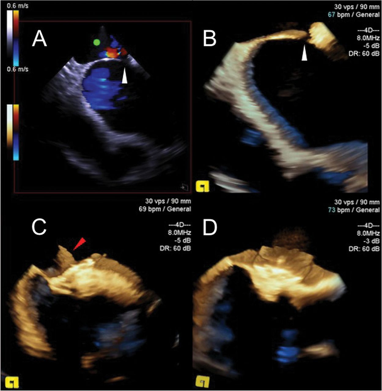
We report the first use of RT3D ICE for PFO closure. A 50-year-old woman underwent PFO closure via the right femoral vein. An ACUSON AcuNav 3D ultrasound catheter (Siemens AG, Germany) inserted via a second right femoral vein puncture provided RT3D ICE images to guide the procedure. RT3D ICE (figure 1B) allowed enhanced anatomical characterisation of the defect (white arrowheads) in comparison with 2D ICE (figure 1A), and reduced ultrasound catheter manipulation during the procedure. The PFO was closed using a 25 mm Ultrasept device (Cardia, Belgium). RT3D ICE images of the device attached to the delivery catheter immediately prior to release (figure 1C) and following successful deployment (figure 1D) are shown. Online supplementary videos 1 and 2 illustrate preclosure and postclosure 2D and RT3D ICE images.
Intracardiac echocardiography (ICE) images of patent foramen ovale (PFO) closure. Panels A and B: two-dimensional (A) and three-dimensional (B) ICE images of the PFO prior to closure (white arrowheads). Panels C and D: three-dimensional ICE images of the PFO closure device straddling the defect before (C) and after (D) release from the delivery catheter (red arrowhead).
Supplementary materials
Supplementary Data
This web only file has been produced by the BMJ Publishing Group from an electronic file supplied by the author(s) and has not been edited for content.
Files in this Data Supplement:
- Data supplement 1 - Online video 1
- Data supplement 2 - Online video 2
Footnotes
-
Contributors CC: specialist registrar who assisted VSM in the PFO closure procedure and wrote the first draft of the manuscript. SAH: cardiac physiologist who obtained and processed the intracardiac echocardiography images during the procedure, and prepared the images for the manuscript figure. VSM: consultant who performed the PFO closure procedure, suggested the images for publication, and revised the first draft into its final version for submission. VSM is responsible for the overall content as guarantor.
-
Competing interests None.
-
Patient consent Obtained.
-
Ethics approval This is a clinical case report and therefore does not require ethics committee approval.
-
Provenance and peer review Not commissioned; internally peer reviewed.