Article Text

Abstract
Since its recognition in December 2019, covid-19 has rapidly spread globally causing a pandemic. Pre-existing comorbidities such as hypertension, diabetes, and cardiovascular disease are associated with a greater severity and higher fatality rate of covid-19. Furthermore, COVID-19 contributes to cardiovascular complications, including acute myocardial injury as a result of acute coronary syndrome, myocarditis, stress-cardiomyopathy, arrhythmias, cardiogenic shock, and cardiac arrest. The cardiovascular interactions of COVID-19 have similarities to that of severe acute respiratory syndrome, Middle East respiratory syndrome and influenza. Specific cardiovascular considerations are also necessary in supportive treatment with anticoagulation, the continued use of renin-angiotensin-aldosterone system inhibitors, arrhythmia monitoring, immunosuppression or modulation, and mechanical circulatory support.
- systemic inflammatory diseases
- cardiac risk factors and prevention
- myocarditis
This is an open access article distributed in accordance with the Creative Commons Attribution Non Commercial (CC BY-NC 4.0) license, which permits others to distribute, remix, adapt, build upon this work non-commercially, and license their derivative works on different terms, provided the original work is properly cited, appropriate credit is given, any changes made indicated, and the use is non-commercial. See: http://creativecommons.org/licenses/by-nc/4.0/.
Statistics from Altmetric.com
Introduction
Severe acute respiratory syndrome coronavirus 2 (SARS-CoV-2), which causes covid-19, was first reported to WHO as a pneumonia of unknown cause in Wuhan, China, on 31 December 2019.1 While the initial outbreak was mostly confined to the epicentre in China, SARS-CoV-2 quickly spreads internationally causing a global pandemic. By 15 March, the number of cases outside of China surpassed those in China, and as of 12 April 2020, over 1.8 million cases and 110 000 deaths were reported worldwide, affecting 185 countries.2
The most common symptoms of COVID-19 include fever, dry cough, myalgia or fatigue, as is the case in many other viral infections.3 4 According to a study by the Chinese Center for Disease Control and Prevention of 44 672 laboratory confirmed, 10 567 clinically diagnosed and 16 186 suspected cases of COVID-19, 81.4% exhibited mild illness (with no or mild symptoms of pneumonia), 13.9% had severe symptoms (dyspnoea with respiratory rate ≥30/min, SpO2 ≤93%, PaO2/FiO2<300 and/or infiltration of lung field >50% within 24–48 hours), and 4.7% were critically ill (respiratory failure, septic shock and/or multiorgan failure).5 In patients with severe or critical disease, viral pneumonia can progress to acute respiratory distress syndrome (ARDS), and multisystem failure accompanied by a cytokine storm.5
Since patients with covid-19 with cardiovascular comorbidities have higher mortality,5 and the severity of COVID-19 disease correlates with cardiovascular manifestations,6 it is important to understand the interaction of COVID-19 and cardiovascular disease (CVD). This review will summarise our current understanding of the cardiovascular manifestations of covid-19, as compared with SARS (caused by SARS-CoV), the Middle East respiratory syndrome (MERS) (caused by MERS-CoV) and influenza. We will also discuss cardiovascular considerations regarding treatment strategies.
Cardiovascular comorbidities
The prevalence of diabetes mellitus (DM) and obesity in patients with COVID-19 in the USA appear to be higher than in the general population, but the prevalence of CVD appears to be similar (table 1). However, in hospitalised patients with COVID-19 with more severe disease, DM, hypertension and CVD seem to be more prevalent in both the USA and China, similar to MERS and influenza (table 2). A retrospective, single-centre case series of 187 patients with COVID-19 found that patients with underlying CVD were more likely to have cardiac injury (troponin (Tn) elevation) compared with patients without CVD (54.5% vs 13.2%).6 In-hospital mortality was 7.6% for patients without underlying CVD and normal Tn, 13.3% for those with CVD and normal Tn, 37.5% for those without CVD but elevated Tn and 69.4% for those with CVD and elevated Tn.6 These observations suggest that although patients with pre-existing CVD may not be more susceptible to contracting SARS-CoV-2, they are prone to more severe complications of COVID-19 with increased mortality.7 The association of cardiovascular and other comorbidities with case fatality rate is summarised in figure 1.

Influence of cardiovascular and other comorbidities on CFR from respiratory viral infections. CDC, Center for Disease Control and Prevention; CFR, case fatality rate; MERS, Middle East respiratory syndrome; SARS, severe acute respiratory syndrome. Data are from Chinese CDC,5 Mertz et al,71 Chan et al 72 and Badawi et al. 73
Prevalence of cardiopulmonary comorbidities in covid-19 disease in the USA and China compared with the general population
Prevalence of comorbidities in hospitalised patients with severe respiratory viral infections
The higher mortality in patients with cardiovascular comorbidities may be directly attributable to underlying CVD or merely coincident with CVD. CVD may also be a marker of accelerated ageing, immune system dysregulation, or other mechanisms that affect COVID-19 prognosis. Age is both a risk factor for CVD and an important determinant of COVID-19 fatality, which has been found to be 14.8% in patients older than 80 years of age but <4% in patients younger than 70 years of age.5 Patients with elevated Tn levels were older than patients with normal Tn (71.4±9.4 vs 53.5±13.2 years).6 In addition, ageing weakens the immune system, as demonstrated by the lower protective titres after influenza vaccination in 50% of adults older than 65 years of age.8
Pathophysiology of cardiac injury
SARS-CoV-2 is an enveloped, positive-sense single-stranded RNA virus.9 SARS-CoV-2 and other similar coronaviruses use the ACE 2 (ACE2) protein for ligand binding before entering the cell via receptor-mediated endocytosis.10 Recent data on viral structure reveal that SARS-CoV-2 has tighter interaction with the human ACE2 receptor binding domain as compared with SARS-CoV, which may explain in part the greater transmissibility of the current virus among humans.11 ACE2 is a membrane protein that serves many physiological functions in the lungs, heart, kidneys and other organs.12 It is highly expressed in type 2 lung alveolar cells, which provides an explanation for the respiratory symptoms experienced by patients with COVID-19.13 More than 7.5% of myocardial cells have positive ACE2 expression, based on single-cell RNA sequencing,14 which could mediate SARS-CoV-2 entry into cardiomyocytes and cause direct cardiotoxicity.
The mechanisms of cardiovascular injury from COVID-19 have not been fully elucidated and are likely multifactorial. SARS-CoV-2 viral particles have been identified by RT-PCR in cardiac tissue in some cases,15 supporting that direct cardiotoxicity may occur. Virally driven hyperinflammation with cytokine release may lead to vascular inflammation, plaque instability, myocardial inflammation, a hypercoagulable state, and direct myocardial suppression.16 17 Other systemic consequences of COVID-19 infection, including sepsis and disseminated intravascular coagulation (DIC), may also mediate cardiac injury. Based on postmortem biopsies, the pathological features of covid-19 in multiple organs greatly resemble those seen in SARS18 19 and MERS.20 21 In cardiac tissue, pathological findings vary from minimal change to interstitial inflammatory infiltration and myocyte necrosis (table 3). In the vasculature, microthrombosis and vascular inflammation could be found (table 3).
Pathological findings of the cardiopulmonary systems in death related to coronaviruses
Cardiovascular manifestations
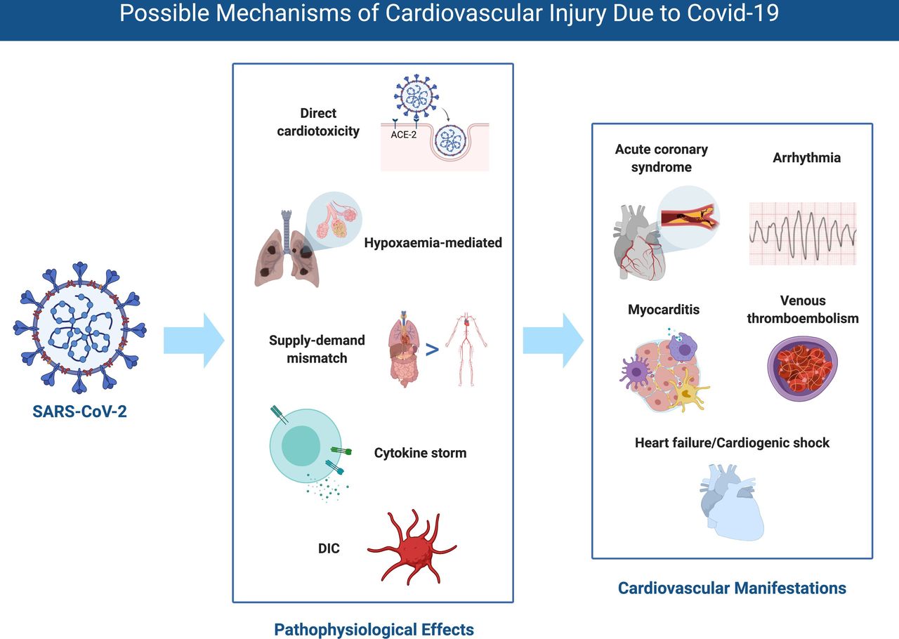
The clinical cardiovascular manifestations of COVID-19 include elevation of cardiac biomarkers (ischaemic or non-ischaemic aetiology), cardiac arrhythmia, arterial and venous thromboembolism (VTE), and cardiogenic shock and arrest. The possible mechanisms and cardiovascular manifestations are shown in figure 2.

Possible mechanisms of cardiovascular injury due to COVID-19. DIC, disseminated intravascular coagulation; SARS-CoV-2, severe acute respiratory syndrome coronavirus 2.
Elevation of cardiac biomarkers
Myocardial injury is common among patients with COVID-19 infection and correlates with disease severity. Although somewhat variable, studies on patients with COVID-19 have generally defined myocardial injury as the elevation of high-sensitivity cardiac troponin (hs-cTn) above the 99th percentile of its upper limit of normal or evidence of new electrocardiographic or echocardiographic abnormalities.3 22 Increased levels of hs-cTn correlate with disease severity and mortality rate in COVID-19, even after controlling for other comorbidities.23 A summary of studies with prevalence of myocardial injury and risk of severe disease or mortality is shown in table 4.
Studies on troponin elevation and risk of severe disease or death in COVID-19
The pattern in the rise of cTn levels is also significant from a prognostic standpoint. Non-survivors had a higher level of Tn elevation which continued to rise until death (mean time from symptom onset to death was 18.5 days (IQR 15–20 days)), while Tn levels for survivors remained unchanged.22 This finding may support the monitoring of cTn levels every few days in hospitalised patients.
It is a challenging task to differentiate potential aetiologies of cardiac injury: acute coronary syndrome (ACS) due to plaque rupture or thrombosis (type I myocardial infarction (MI)) or supply-demand mismatch (type II MI), myocardial injury due to DIC, and non-ischaemic injury (myocarditis, stress-induced cardiomyopathy, or cytokine release syndrome).
Ischaemic myocardial injury
Myocardial infarction
Severe viral infections can cause a systemic inflammatory response syndrome that increases the risk of plaque rupture and thrombus formation, resulting in either an ST-elevation MI or non-ST-elevation MI.24 In a study of 75 patients hospitalised with SARS, acute MI was the cause of death in two of five fatal cases.25 There is also a significant association between acute MI and influenza. As compared with the prevalence of acute MI occurred 1 year before or 7 days to 1 year after influenza, the incidence ratio of acute MI within 7 days of influenza infection was 6.1 (95% CI: 3.9 to 9.5).26 Although reports of type I MI in patients with COVID-19 have not yet been published, anecdotal report was presented.27 Treatment of ACS in COVID-19 should be according to the updated Society for Cardiovascular Angiography and Interventions guidelines.28
Severe respiratory viral infections can also lead to decreased oxygen delivery to the myocardium via hypoxaemia and vasoconstriction, as well as the haemodynamic effects of sepsis with increased myocardial oxygen demand. This supply and demand mismatch may lead to sustained myocardial ischaemia in patients with underlying coronary artery disease. However, a rise and/or fall of hs-cTn is not sufficient to secure the diagnosis of acute MI as seen in MI with non-obstructive coronaries, even in the absence of COVID-19. Therefore, the diagnosis of acute MI should also be based on clinical judgement, symptom/signs, ECG changes, and imaging studies.
Myocardial injury with DIC
DIC is a life-threatening condition present in 71.4% (15/21) of non-survivors with COVID-19 and 0.6% (1/162) of survivors.29 A marker of severe sepsis, DIC further perpetuates multiorgan damage through thrombosis, reduced perfusion, and bleeding, DIC has been implicated in the thrombosis of coronary arteries (epicardial vessels and microvasculature), focal necrosis of the myocardium, and severe cardiac dysfunction.30 Myocardial injury with DIC has been recently reported in two critically ill patients with COVID-19.31 Both patients had significantly elevated Tn and brain natriuretic peptide, which normalised after treatment with heparin, mechanical ventilation, and antiviral agents.
Non-ischaemic myocardial injury
Myocarditis and stress-induced cardiomyopathy
Myocardial injury from SARS-CoV-2 infection may also be mediated by non-ischaemic mechanisms, such as acute and fulminant myocarditis and stress-induced cardiomyopathy. Reports of various presentations of non-ischaemic myocardial injury in COVID-19 are summarised in table 5. The distinction between myocarditis and stress-induced cardiomyopathy can be challenging, since cardiovascular magnetic resonance (CMR) and/or biopsy are not available in most cases. Fried et al and Sala et al each reported a patient with COVID-19 with mid-left ventricular (LV) or basal-to-mid LV hypokinesis, a pattern of mid-ventricular, or reverse Takotsubo stress cardiomyopathy, respectively.32 33 The incidence of acute heart failure was 33% (7/21) in critically ill patients with COVID-19 without a past history of LV systolic dysfunction in Washington state.34 Importantly, cardiomyopathy can develop in COVID-19 with mild or absent respiratory symptoms.35
Clinical characteristics of non-ischaemic myocardial injury in covid-19
Myocardial injury with cytokine release syndrome
Similar to SARS-CoV and MERS-CoV, SARS-CoV-2 can elicit the intense release of multiple cytokines and chemokines by the immune system.3 36 Cytokine release syndrome (aka ‘cytokine storm’), a poorly understood immunopathological process caused by hyperinduction of proinflammatory cytokines such as interleukin (IL)-1, IL-6, T helper 1 cytokine interferon-gamma, and tumour necrosis factor-alpha (TNF-α), has been reported in the setting of SARS, MERS, and influenza.37–39 It is postulated that proinflammatory cytokines depress myocardial function immediately through activation of the neural sphingomyelinase pathway and subacutely (hours to days) via nitric oxide-mediated blunting of beta-adrenergic signalling.40 Accumulating evidence suggest that a subgroup of patients with severe COVID-19 can develop cytokine storm.36 Plasma levels of IL-1β, IL-6, IL-8 and TNF-α have been found to be significantly higher in patients with COVID-19.3 The clinical and biochemical profiles of non-survivors in patients with COVID-19 with highly elevated ferritin and IL-6 also suggest that cytokine release contribute to mortality.41
Arrhythmia
Arrhythmia could be the first presentation of COVID-19, and new-onset and/or progressive arrhythmia could indicate cardiac involvement. A study of 137 patients in Wuhan showed that 7.3% had experienced palpitations as one of their presenting symptoms for COVID-19.42 The prevalence of unspecified arrhythmia in two additional studies is listed in table 4. Arrhythmias were found to be more common in intensive care unit (ICU) patients with COVID-19 (44.4%) than non-ICU patients (6.9%).4 Patients with elevated Tn also had a higher incidence of malignant arrhythmia (haemodynamically unstable ventricular tachycardia or ventricular fibrillation) than those with normal Tn levels (11.5% vs 5.2%, p<0.001).6
Venous thromboembolism
Due to prolonged immobilisation, hypercoagulable status, active inflammation, and propensity for DIC, patients with COVID-19 are at increased risk of VTE. The prevalence of ultrasound-confirmed deep venous thrombosis in patients with COVID-19 is 22.7%43 and 27% in ICU patients.44 Patients with cCOVID-19 have been shown to have significant higher level of D-dimer, fibrin degradation products (FDP), and fibrinogen, compared with healthy controls.45 In addition, D-dimer and FDP titres were higher in patients with severe COVID-19 than those with milder disease.45 A retrospective, multicentre cohort study demonstrated that D-dimer >1 µg/mL on admission was associated with in-hospital death (OR: 18.4, 95% CI: 2.6 to 128.6, p=0.003).22 Thus, in the setting of critically ill COVID-19 patients with clinical deterioration, VTE (including pulmonary embolism) should be considered.
Treatment considerations
There is currently no proven therapy for COVID-19, although many are under investigation. We focus our discussion here on supportive therapies and considerations of potential therapies relevant to the cardiovascular system, summarized in table 6.
Cardiovascular considerations in treatment
Anticoagulant therapy
Due to the high rate of associated arterial thromboembolism and VTE, prophylactic anticoagulation is essential in the management of hospitalised patients with COVID-19,44 46 although the optimal thromboprophylaxis regimen is unclear. In a retrospective study of 449 patients with severe COVID-19, 99 patients received unfractionated heparin or low molecular weight heparin for at least 7 days.47 No difference in overall 28-day mortality was observed, but in subgroups of patients with sepsis-induced coagulopathy score ≥4, or D-dimer >sixfold of upper limit of normal, the heparin group had lower mortality compared with the no-heparin group (40.0% vs 64.2%, p=0.029), (32.8% vs 52.4%, p=0.017), respectively.47 Another concern regarding thromboprophylaxis is the drug-drug interaction between some antiviral treatments (such as ribavirin, lopinavir and ritonavir) and direct oral anticoagulants. Low molecular weight heparin is likely preferred in critically ill patients with COVID-19, if anticoagulation is used.
ACE inhibitors and angiotensin receptor blockers
Since SARS-CoV-2 uses the ACE2 protein for ligand binding before entering the cell,10 there are concerns regarding the use of renin-angiotensin-aldosterone system (RAAS) inhibitors that may increase ACE2 expression.13 48 The relationship between ACE2 expression and SARS-CoV-2 virulence is uncertain. After cell entry via ACE2, SARS-CoV-2 appears to subsequently downregulate ACE2 expression, which is vital to maintenance of cardiac function.49 In mouse models, reduction in ACE2 expression is associated with increased severity of lung injury induced by influenza.50 Whether higher expression of ACE2 increases susceptibility to SARS-CoV-2 infection or confers cardioprotection remains controversial.51 Furthermore, experimental animal models and few studies in humans have yielded conflicting results as to whether RAAS inhibition increases ACE2 expression.52 53 Based on the uncertainty regarding the overall effect of RAAS inhibitors (ACE inhibitor (ACEI) and angiotensin receptor blocker (ARB) therapy) in COVID-19, multiple specialty societies currently recommend that RAAS inhibitors be continued in patients in otherwise stable condition.51
Hydroxychloroquine/Chloroquine and azithromycin
Chloroquine is a versatile bioactive agent with antiviral activity in vitro against DNA viruses as well as various RNA viruses, including SARS-CoV, MERS-CoV, and SARS-CoV-2, prompting it to be studied for patients with COVID-19.54 Similar to chloroquine, hydroxychloroquine confers antiviral effects and has an additional modulating effect on activated immune cells to decrease IL-6 expression.55 Non-randomised, observational studies of >100 patients at 10 hospitals in China suggest that chloroquine or hydroxychloroquine is superior to the control treatment in resolving pneumonia (based on clinical and imaging findings), promoting conversion to viral negative status and shortening the disease course.56 In a small open-label, non-randomised study conducted in France, 70% of hydroxychloroquine-treated patients with COVID-19 had undetectable viral titres at 6 days, compared with 12.5% in the control group (p=0.001),57 although there were major methodological concerns with the study. Azithromycin, a macrolide antibiotic which acts against Zika and Ebola viruses in vitro58 and suppresses inflammatory processes,59 has been proposed as an effective adjunct to hydroxychloroquine in COVID-19 through unclear mechanisms.57 Although the preliminary evidence for quinoline antimalarials may be promising, it should be taken with caution until randomised clinical trials are completed.
Both chloroquine and azithromycin have generally favourable safety profiles, but they are known to cause cardiovascular side effects including prolongation of QT interval.60 Although azithromycin interferes minimally with the cytochrome P-450 system in vitro,61 chloroquine is metabolised by CYP2C8 and CYP3A4/5,62 and there is a potentially enhanced risk of significant QT prolongation induced by combination therapy. Guidance for managing QTc prolongation in COVID-19 pharmacotherapies and other cardiac electrophysiology issues related to covid-19 has recently been published by the Heart Rhythm Society.63
Immunosuppressive therapy
As for SARS and MERS, corticosteroids are not routinely recommended for COVID-19 and might exacerbate the resulting lung injury.64 However, given the detrimental effects of the cytokine release syndrome associated with COVID-19 on the cardiopulmonary system, immunosuppressive therapy to dampen the hyperinflammatory response may be beneficial.36 Screening patients with COVID-19 using laboratory data (increasing ferritin and erythrocyte sedimentation rate, and decreasing platelets) and a proposed score for the hemophagocytic response (HScore)65 for hyperinflammation may help to identify patients for whom immunosuppression might improve survival.36
IL-6 inhibitors may have a role as immunomodulators. Tocilizumab, an inhibitor of the IL-6 receptor, was shown to effectively improve symptoms and prevent clinical deterioration in a retrospective case series study of 21 COVID-19 patients with severe or critical disease.66 Several clinical trials are ongoing to assess the efficacy and safety of tocilizumab (NCT04317092)67 or sarilumab, another IL-6 inhibitor (NCT04327388)68 in patients with COVID-19.
Mechanical cardiopulmonary support
Mechanical cardiopulmonary support in respiratory failure has been reported with variable survival rates. Extracorporeal Life Support Organization Registry is being adapted to investigate COVID-19 in prospective observational studies addressing the role of extracorporeal membrane oxygenation (ECMO) in these critically ill patients.69
In the setting of cardiogenic shock related to COVID-19, intra-aortic balloon pump (IABP), or veno-arterial ECMO should be considered. During the 2009 H1N1 pandemic in Japan, 9 out of 13 patients with fulminant myocarditis receiving emergent mechanical circulatory support (IABP and/or percutaneous cardiopulmonary support) survived, while all four patients with fulminant myocarditis treated without mechanical circulatory support died.70 A case has been reported of a patient with COVID-19 without respiratory symptoms presenting with cardiogenic shock attributed to acute myopericarditis and successfully treated with IABP support.32 Perhaps a more likely scenario is the conversion of veno-venous ECMO for ARDS to veno-arterial-venous ECMO on development of cardiogenic shock, as described in another case report.32 Regardless of the mode of mechanical support, patient selection should involve consideration of comorbidities and potential complications.
Conclusion
COVID-19 is similar to SARS and MERS with regard to host vulnerability, specifically in those with substantial cardiovascular comorbidities. Greater transmissibility of COVID-19 has resulted in a worldwide pandemic, a record number of infected individuals and an excess mortality that far exceeded previous coronavirus-related outbreaks. Myocardial injury is common in COVID-19 and portends a worse prognosis. Differentiating between the various causes of myocardial injury is crucial to determining the treatment course. The cardiovascular considerations for treatment, including anticoagulation, ACEI or ARB use, anti-arrhythmic management, immunosuppression/modulation, and haemodynamic support, are important and continue to evolve.
References
Footnotes
Twitter @tiffchenMD
Contributors YK, TC, DM, and YH drafted the manuscript. All authors critically reviewed and edited the manuscript.
Funding The authors have not declared a specific grant for this research from any funding agency in the public, commercial or not-for-profit sectors.
Competing interests None declared.
Patient and public involvement Patients and/or the public were not involved in the design, conduct, reporting or dissemination plans of this research.
Patient consent for publication Not required.
Provenance and peer review Not commissioned; externally peer reviewed.