Article Text

Abstract
Background: The mechanisms of progression from fatty liver to steatohepatitis and cirrhosis are not well elucidated. Mitochondrial dysfunction represents a key factor in the progression of non-alcoholic steatohepatitis (NASH) as mitochondria are the main cellular site of fatty acid oxidation, ATP synthesis and reactive oxygen species (ROS) production.
Aims: (1) To evaluate the role of the uncoupling protein 2 in controlling mitochondrial proton leak and ROS production in NASH rats and humans; and (2) to assess the acute liver damage induced by ischaemia–reperfusion in rats with NASH.
Methods: Mitochondria were extracted from the livers of NASH humans and rats fed a methionine and choline deficient diet. Proton leak, H2O2 synthesis, reduced glutathione/oxidised glutathione, 4-hydroxy-2-nonenal (HNE)–protein adducts, uncoupling protein-2 (UCP2) expression and ATP homeostasis were evaluated before and after ischaemia–reperfusion injury.
Results: NASH mitochondria exhibited an increased rate of proton leak due to upregulation of UCP2. These results correlated with increased production of mitochondrial hydrogen peroxide and HNE–protein adducts, and decreased hepatic ATP content that was not dependent on mitochondrial ATPase dysfunction. The application of an ischaemia–reperfusion protocol to these livers strongly depleted hepatic ATP stores, significantly increased mitochondrial ROS production and impaired ATPase activity. Livers from patients with NASH exhibited UCP2 over-expression and mitochondrial oxidative stress.
Conclusions: Upregulation of UCP2 in human and rat NASH liver induces mitochondrial uncoupling, lowers the redox pressure on the mitochondrial respiratory chain and acts as a protective mechanism against damage progression but compromises the liver capacity to respond to additional acute energy demands, such as ischaemia–reperfusion. These findings suggest that UCP2-dependent mitochondria uncoupling is an important factor underlying events leading to NASH and cirrhosis.
Statistics from Altmetric.com
Non-alcoholic fatty liver disease (NAFLD) identifies a pathological condition characterised by histological features of alcoholic liver disease that occur in subjects with minimal or no alcohol intake.1 Non-alcoholic steatohepatitis (NASH) specifically refers to that subset of NAFLD patients with evidence of inflammation on liver biopsy. The prevalence of NASH in Western countries is around 2 to 3%,2 and is currently considered a significant cause of cryptogenic cirrhosis3 and liver transplantation.4
Although the pathogenesis of NASH is not completely understood, a growing body of evidence suggests that the development of NASH requires a double “hit”:5 the first “hit” involves the accumulation of fatty acids in the liver, and the second includes oxidative stress,6 decreased hepatic ATP production7 and induction of pro-inflammatory cytokines.8 Mitochondria play a key role in the development of NASH, as the main cellular site of fatty acid oxidation, ATP synthesis and production of reactive oxygen species (ROS). During steatosis, hepatocytes are supplied by large amounts of fatty acids, which increase mitochondrial respiratory activity,9 induce increased ROS production and promote liver oxidative stress. NASH patients present mitochondrial abnormalities10 and increased ROS production.11 12 Uncoupling protein-2 (UCP2) is a mitochondrial inner membrane protein with ubiquitous tissue distribution, mediating proton leak across the inner membrane by uncoupling substrates oxidation from ATP synthesis.13 In physiological conditions UCP2 is expressed in hepatic non-parenchymal cells14 but not in hepatocytes. We hypothesised that during NASH progression, the upregulation of UCP2 acts as protective mechanism against excessive fat accumulation but exposes the liver to chronic depletion of ATP, which increases the susceptibility of the hepatocytes to conditions of acute energy demand, ie, ischaemia–reperfusion injury. We also demonstrated that UCP2 is over-activated by mitochondrial membrane lipoperoxidation, suggesting a strength modulation along the mitochondrial respiratory chain, fat storage, ROS production and UCP2 during NASH progression.
Dietary-induced deficiency of choline and methionine was chosen, from 3 to 11 weeks of the disease, to study the progression of NASH since it is currently considered the model that better imitates the change in redox balance observed in NAFLD.15–17
The clinical relevance of the hypothesis suggested in the present study was also supported by our observation of UCP2 over-expression, 4-hydroxy-2-nonenal (HNE) increase and mitochondrial oxidative stress in human liver during the development of NASH.
MATERIALS AND METHODS
Animal experiments
Male Wistar rats (Harlan, S. Pietro al Natisone, UD, Italy) weighing 350–400 g were caged individually in a temperature/light controlled environment with free access to food and water. All rats received care in compliance with the Principles of Laboratory Animal Care formulated by the National Society for Medical Research and the Guide for the Care and Use of Laboratory Animals prepared by the Institute of Laboratory Animal Resources (NIH Publication No. 86–23, revised 1985).
Steatohepatitis was induced by a high fat/methionine and choline deficient diet (MCD). Control rats consumed the same diet with dl-methionine (3 g/kg) and choline bitartrate (2 g/kg) supplementation. Both diets were purchased from Mucedola (Settimo Milanese, MI, Italy).18 At the end of 3, 7 and 11 weeks of diet, five rats for each time were anaesthetised by i.p. 100 mg/kg ketamine and 2.5 mg/kg acepromazine and then sacrificed, their blood drawn and livers removed.
Liver ischaemia–reperfusion
The response to a sublethal, acute stress (eg, hepatic ischaemia–reperfusion) was studied in control (n = 7) and NASH rats at 3 and 11 weeks of diet (n = 7 for each time) using a Schwartz’s clip for 60 min as previously reported.19 Twenty-four hours after reperfusion, animals were sacrificed and blood and livers processed. Steatohepatitis was confirmed by serum alanine aminotransferase (ALT) activity, histological analysis and collagen III expression.
Human study
Liver specimens were obtained during non-hepatic abdominal surgery from 10 consecutive patients (age, 39–60 years; seven women) affected by non-alcoholic steatohepatitis (NASH group), defined by histology; and eight subjects (age 35–69 years, six women) each with normal liver (healthy group).
All subjects included in the study were negative for viral hepatitis infection, liver autoimmune or metabolic disorders and were not under treatment with hepatotoxic drugs. All patients gave written informed consent to the study.
Preparation of cells and isolation of mitochondria
Liver cell suspensions were prepared from rats by using perfusion with collagenase as previously described.20 The suspension obtained was filtered through nylon gauze (mesh width, 50 μm) and centrifuged for 2 min at 50 g at 4°C. This centrifugation resulted in a pellet enriched in parenchymal cells and a supernatant of non-parenchymal cells, which was discarded. Parenchymal cells were then purified in Percoll (1.08 g/ml of density) to eliminate damaged parenchymal cells and residual non-parenchymal cells.21
Mitochondria were isolated from human and rat livers as previously described.22 Protein concentration was determined using the Lowry micromethod kit (Sigma–Aldrich, St. Louis, Missouri, USA).
Biochemical analysis
Determination of mitochondrial inner membrane potential and oxygraphic measurements
Freshly prepared mitochondria were assayed for mitochondrial membrane potential (Δψ) at 37°C, in the presence of succinate, rotenone and oligomycin by a Clark’s electrode and a tetraphenylphosphonium (TPP+) electrode (WPI, Berlin, Germany). TPP+ uptake was carried out in basal conditions and in the presence of guanosine diphosphate (GDP), a potent and specific inhibitor of UCPs. Membrane potential calculations were made using a modified Nernst equation as previously reported.22 The determination of the dependence of membrane potential of the proton leak activity in isolated mitochondria is based on the protocol described by Porter and Brand.23
Evaluation of F0F1-ATPase activity and hepatic ATP content
F0F1-ATPase activity was measured following ATP hydrolysis with an ATP-regenerating system coupled to oxidation of the reduced form of nicotinamide–adenine dinucleotide phosphate (NADPH).24 Liver ATP content was measured using the method of Yang et al25 and the concentration was assessed by a commercial bioluminescence assay kit (Sigma–Aldrich).
Measurement of mitochondrial H2O2 production
The rate of peroxide production was determined in isolated liver mitochondria by a modification of the method of Barja, as previously reported.20
Glutathione assay
Mitochondrial levels of reduced glutathione (GSH) and oxidised glutathione (GSSG) were measured by high-performance liquid chromatography (HPLC) according to Viña, as described previously.20
Mitochondrial 4-hydroxy-2-nonenal–protein adducts
Fluorescent adducts formed between 4-hydroxy-2-nonenal (HNE) and mitochondrial proteins were monitored by spectrofluorimetry as previously reported26 in human and rat livers.
Analysis of protein and mRNA expression
Immunoprecipitation and western blot
Mitochondrial proteins (500 μg) isolated from parenchymal liver cells were immunoprecipitated at 4°C overnight with 0.4 μg anti-UCP2 antibody (rabbit polyclonal UCP2 from Santa Cruz Biotechnology, Santa Cruz, California, USA) and 50 μl protein G-agarose beads, using the Protein G Immunoprecipitation Kit (IP-50; Sigma–Aldrich). The immunoprecipitated proteins were loaded onto 12% SDS-PAGE gels and transferred to nitrocellulose membrane for immunoblot analysis with anti-UCP2 antibodies. The membranes were then stripped by Restore Stripping Buffer (#21059; Pierce, Rockford, Illinois, USA) for 30 min, washed three times and incubated with anti-HNE antibodies (1:1000; Alpha Diagnostic, San Antonio, Texas, USA) overnight. Secondary antibodies were conjugated with horseradish peroxidase and immunoblots detected by VersaDoc Image System (Bio-Rad Laboratories, Hercules, California, USA) as previously reported.27
RNA extraction and real-time reverse transcriptase polymerase chain reaction
Liver tissues stored at −80°C were homogenised and total RNA was isolated using the RNeasy kit (QIAGEN GmbH, Hilden, Germany) as previously reported.20 cDNA was obtained using a random hexamer primer and a SuperScript III Reverse Transcriptase kit as described by the manufacturer (Invitrogen, Frederick, MD, USA). A PCR master mix containing the specific primers (rat UCP2: forward, 5′-CTT TGA AGA ACG GGA CAC-3′; reverse, 5′-TCC TGC TAC CTC CCA GGA-3′; human UCP2: forward, 5′-TTG AAG AAC GGG ACA CCT TT-3′; reverse, 5′-GGG CAC CTT TAA TCA GCA AC-3′; collagen III: forward, 5′-TTC TGC CAC CCT GAA CTC-3′; reverse, 5′-ATC TGT CCA CCA GTG CTT C-3′; rat glyceraldehyde-3-phosphate dehydrogenase (GAPDH): forward, 5′-TCA AGG CTG AGA ATG GGA AG-3′; reverse: 5′-ATG GTG GTG AAG ATG CCA GT-3′; human GAPDH: forward, 5′-CAA GGC TGA GAA CGG GAA-3′; reverse: 5′-GCA TCG CCC CAC TTG ATT TT-3′) was added, along with AmpliTaq Gold DNA polymerase (Applied Biosystems, Foster City, CA, USA). Real-time quantification of mRNA was performed with a SYBR Green I assay, and evaluated using an iCycler detection system (Bio-Rad Laboratories). The threshold cycle (CT) was determined, and the relative genes expression subsequently was calculated as follows: fold change = 2−Δ(ΔCT), where ΔCT = CTtarget − CThousekeeping and Δ(ΔCT) = ΔCTtreated − ΔCTcontrol.
Statistical analysis
Data were expressed as mean ± standard deviation of the mean (SDM). Since the data were not paired, differences among means were analysed using one-way ANOVA after Gaussian distribution evaluation by the Kolgomorov–Smirnov test. The Tukey–Kramer multiple comparison test for all pairs of columns was applied as a post hoc test. In all instances p<0.05 was taken as the lowest level of significance. The package GraphPad Prism 4 for Windows was used to perform all the statistical analyses.
RESULTS
Induction of NASH and liver pathology
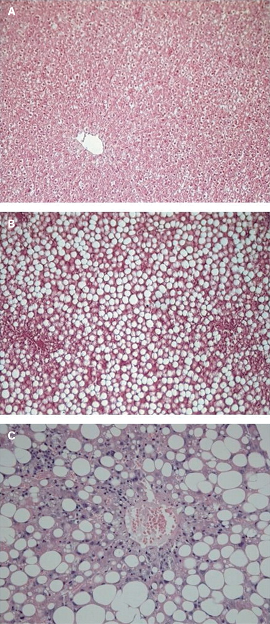
ALT levels significantly increased at 3 weeks as compared to control rats (p<0.01) and persisted at high levels at 7 and 11 weeks (p<0.01 vs controls, fig 1A). Expression of collagen III mRNA was significantly higher at 3 weeks and increased at 7 and 11 weeks of the MCD diet (fig 1B). Histochemical analysis of liver specimens stained with haematoxylin and eosin (H&E) from rats fed the normal or MCD diet for 3 and 11 weeks are reported in fig 2.

Mitochondrial proton leak increased during NASH development
To verify whether changes in mitochondrial membrane potential and permeability occur during NASH development, mitochondria from NASH and control livers were incubated with succinate as substrate and rotenone as complex I inhibitor in the presence of oligomycin, which selectively inhibits mitochondrial ATP synthase (fig 3).

Normal mitochondria exhibited a bi-phasic relationship between the rate of respiration and the extent of the membrane potential (fig 3A). At a relatively low respiratory rate a linear increase of the membrane potential was measured up to around 150 mV. Further increase of the respiratory activity resulted in a lower rate of enhancement of the membrane potential which remained contained within 180 mV. When NASH mitochondria were studied, the profile of the flow–force relationship was different especially at 7 and 11 weeks of MCD diet treatment (fig 3C,D). Notably, at 7 weeks of NASH a significant change in the correlation slope was found resulting in a much higher oxidation rate of succinate required to settle a given membrane potential when compared to the controls.
The explanation for these observations is that an uncoupling agent progressively increases the proton leak during NASH development. In fact, in the presence of GDP, a potent and specific inhibitor of UCP2, this effect was completely abolished in NASH mitochondria whereas there were no changes in controls. These latter results argue against the involvement of decoupling of the respiratory chain proton pumps and/or changes in the intrinsic inner mitochondrial membrane permeability, and alternatively point to a specific contribution of the uncoupling proteins.
UCP2 expression during NASH development
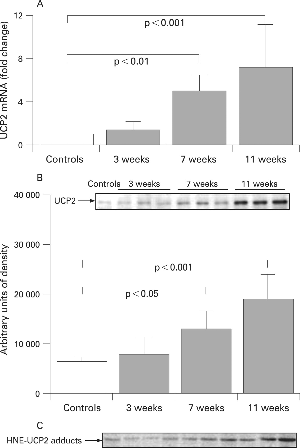
Uncoupling protein-2 (UCP2) is a mitochondrial inner membrane protein that regulates coupling between the electrons transport chain and ATP synthesis. UCP2 expression in healthy liver is primarily limited to Kupffer cells.14 However, UCP2 becomes strikingly abundant in hepatocytes of fatty liver.28 29 Since the role played by parenchymal UCP2 in the progression of NASH is debated, we isolated mitochondrial proteins from NASH and control rat hepatocytes and observed that UCP2 was expressed scantily in normal hepatocytes but was upregulated at 7 and 11 weeks of NASH development (fig 4).
Hepatic ATP depletion occurs during NASH progression but it is not dependent on the F0F1-ATPase alteration
Since UCP2 modulates the coupling between substrate oxidation and ATP synthesis by dissipating the proton motive force used by the complex V for producing ATP, we investigated whether liver ATP stores changed during NASH progression. Hepatic ATP content was constantly lower in NASH than in control rats at 3, 7 and 11 weeks (p<0.001, table 1). Very interestingly, the specific ATPase activity was unaffected in NASH at 3 and 7 weeks as compared to controls and increased at 11 weeks (table 1).
UCP2-dependent proton leak limits ROS production during NASH progression
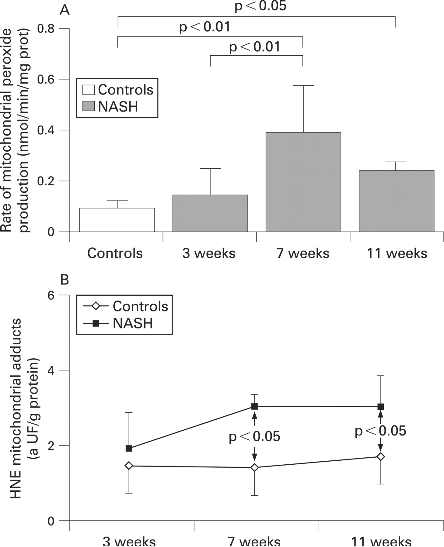
Production of mitochondrial hydroperoxide was measured using succinate as a complex II-linked substrate. The rate of peroxide synthesis significantly increased in NASH mitochondria at 7 and 11 weeks as compared to controls (p<0.01; fig 5A).

We also analysed the level of mitochondria HNE–protein adducts during NASH development. We observed an increase in HNE–protein adducts which became evident at 7 and 11 weeks (p<0.05 at 7 and 11 weeks v controls, fig 5B).
To explain whether HNE can interact with UCP2, we immunoprecipitated mitochondrial proteins with an anti-UCP2 antibody, and revealed the blot with an anti-HNE adduct as reported in fig 4C. Aspecific reactions were observed in the controls and at 3 weeks of NASH. The reactivity of the HNE–UCP2 adducts was evident at 7 and 11 weeks of NASH.
Acute ischaemia–reperfusion injury induced ATP depletion and impairment in ATPase activity in NASH
To verify the hypothesis that UCP2 upregulation may decrease the efficiency of ATP synthesis and compromise the capacity of the liver to respond to acute energy supply, NASH and control rats were subjected to 60 min of liver ischaemia and 24 h of reperfusion at 3 (when UCP2 and proton leak are not yet significant) and 11 weeks.
Ischaemia–reperfusion induced significant liver damage in NASH when compared to control rats subjected to the same protocol, as indicated by the ALT level (fig 6A). Ischaemia–reperfusion injury induced a significant over-production of H2O2 in NASH liver at 11 weeks which was not observed at 3 weeks (fig 6B). The ATP content was also significantly reduced in NASH rats at 11 weeks as compared to 3 weeks (fig 6C). The loss of hepatic ATP stores was only partially justified by the impairment of the ATPase activity, since we observed a dramatic decrease of complex V activity in NASH as well as in control rats subjected to ischaemia–reperfusion (fig 6D).

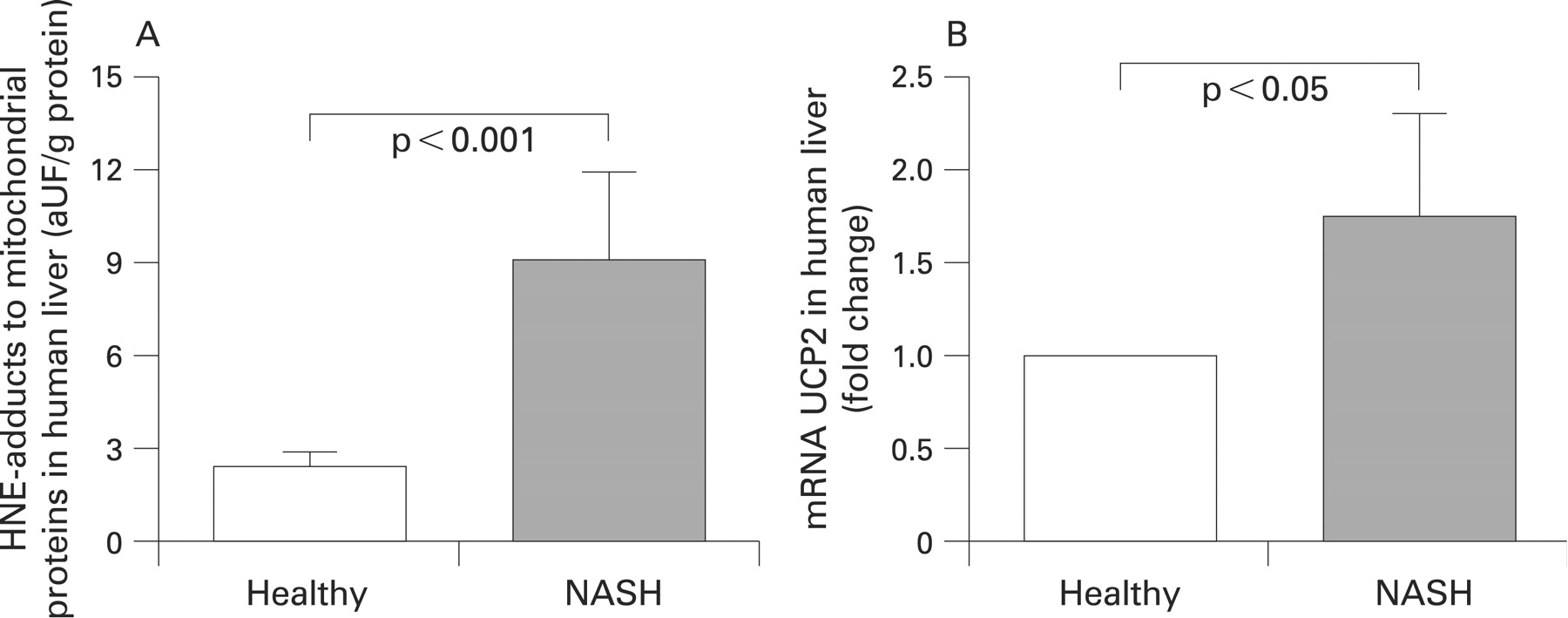
Patients with NASH present mitochondrial oxidative stress, HNE over-production and UCP2 over-expression
To verify whether mitochondrial oxidative stress and UCP2 over-expression occur in human disease, the redox balance was studied in patients affected by NASH and in healthy subjects by measuring mitochondrial GSH and GSSG levels, the protein oxidation by analysis of mitochondrial HNE adducts, and expression of mRNA UCP2 by RT-PCR. Reduced glutathione (GSH) was significantly lower in NASH liver mitochondria as compared with mitochondria from healthy subjects (p<0.001, table 2), whereas the oxidised glutathione (GSSG) significantly increased in NASH (p<0.05, table 2). The concentration of HNE adducts was significantly higher in NASH mitochondria as compared to those from healthy subjects (p<0.001, fig 7A). RT-PCR analysis revealed that UCP2 mRNA was over-expressed by about 2-fold in NASH as compared to normal liver (p<0.05, fig 7B).
DISCUSSION
NASH is characterised by an overload of free fatty acids (FFAs) in hepatocytes that reach a new energetic steady state, whereby the increased hepatic uptake and synthesis of FFAs are compensated by an increased hepatic FFAs removal through β-oxidation.30
Increased electron transport activity in mitochondrial respiratory chain (MRC) of obese mouse liver has been reported,28 suggesting an adaptive response of mitochondria to enhance the capacity of substrates oxidation.9 31 The first aim of the present study was to investigate the possibility that this adaptation occurs primarily during steatosis and its implication for hepatic energy metabolism.
Of interest, we have found that at any time of disease development, the liver ATP content is reduced as compared to controls. These data agree with the results of Cortez-Pinto et al,7 who observed ATP depletion in liver from NASH patients, but our data suggest that the depletion occurs earlier. To verify whether the reduced ATP content was dependent on the impairment of mitochondrial ATP synthesis, we analysed the activity of the F0F1-ATPase but did not find any reduction between NASH and controls. In contrast, ATP-synthase activity increased upon NASH development. This latter result ruled out the possibility that the observed decrease in the steady-state ATP level in NASH liver was due to a reduced content or activity of ATP synthase.
Since during NASH development the substrate supply exceeds the energy requirements, our findings suggest that hepatocytes activate an alternative substrate oxidation pathway not coupled to ATP synthesis.
We demonstrate for the first time that, during NASH development, mitochondria progressively increase the rate of proton leak, which partially dissipates the membrane potential, suggesting the action of an uncoupling agent. UCP2 modulates the coupling between substrates oxidation and ATP synthesis,32 acting as mitochondrial proton carrier and not directly on the F0F1-ATPase.33 In physiological conditions, UCP2 is not expressed in hepatocytes.14 Recent studies have reported an increased expression of UCP2 in hepatocytes of obese liver and that lipids upregulate UCP2 expression in vitro.28 34 Our data show that the increase in proton conductance occurring during NASH progression depends on UCP2 activation. Accordingly, the proton leak was completely reversed by the use of GDP, a potent and specific inhibitor of UCP2.
Hampering electron transfer within the MRC causes over-reduction of some respiratory chain components, which react with oxygen to form the superoxide anion radical,35 which is converted to H2O2 by superoxide dismutase. H2O2 production correlates with membrane potential and both can be reduced by artificial uncouplers.36 Furthermore, small changes of membrane potential have large effects on the rate of superoxide production.37 Pérez-Carreras et al11 reported a defective hepatic MRC in NASH patients. Our data show an increase in mitochondrial H2O2 production in NASH together with UCP2 expression. Nevertheless, mitochondrial H2O2 generation does not increase progressively upon NASH development, as it occurs in other liver diseases such as biliary cirrhosis.20 This may be due to UCP2 upregulation that limits mitochondria oxidative stress in NASH hepatocytes.38
Free radicals oxidise polyunsaturated fatty acyl groups of membrane phospholipids producing various aldehydes such 4-hydroxy-2-nonenal (HNE), a reactive mediator of free-radical damage.39 Low concentrations of HNE may act as a biological signal to induce mitochondrial uncoupling.40
Using human and rat livers we have reported a significant increase in mitochondrial HNE–protein adducts during NASH development, expressed as mitochondrial oxidative stress. Moreover, our data suggest a direct relationship between HNE release and UCP2 activation. Echtay et al40 were the first to suggest that HNE may act as activator of uncoupling, but they limited the observation to normal hepatocytes, which do not express UCP2. In the present work we demonstrated that the increase in HNE–mitochondrial protein adducts was temporally associated with increased proton leak and also with the appearance of HNE–UCP2 adducts. The mitochondrial oxidative stress was also measured in human NASH liver in terms of mitochondrial GSH depletion and GSSG production. The depletion of GSH and increase in GSSG observed in our patients strengthens the idea that mitochondrial oxidative stress may play a key role in the progression of NASH.
In fig 8 we propose a possible mechanism that may explain the link between UCP2, mitochondrial proton leak and modulation of oxidative stress. Induction of UCP2 during NASH progression, by uncoupling oxidative phosphorylation, diminishes the redox pressure on the mitochondrial electron transport chain and provides an added advantage by limiting the production of mitochondrial ROS.41 However, over-expression of UCP2, by decreasing the efficiency of energy synthesis,42 compromises the liver capacity to respond to an acute energy need. Our findings support a recent report by Rizki et al43 who found that 3 weeks of a lipogenic MCD diet in the murine model provoked a substantial (26%) loss of body weight despite an increased metabolic rate. This is in line with the functional features of the UCPs that, by uncoupling the mitochondrial oxidative phosphorylation system, cause increased substrate oxidation without a gain in ATP synthesis.

The MCD model of NASH is the most widely known and, although it is not completely transferable to humans, at 11 weeks the liver showed a significant increase in inflammation infiltrate miming the shift from steatosis to NASH observed in human disease. We have firstly demonstrated an increase in the UCP2 expression in human liver which supports the role of mitochondrial adaptation as an essentially patho-physiological mechanism in the development of NASH.
Our theory is supported by the experiment of acute damage induced by ischaemia–reperfusion injury. NASH liver aggravated ischaemia–reperfusion damage and induced a more dramatic depletion of ATP which was not completely explained by the impairment of ATPase which occurs in all livers subjected to ischaemia–reperfusion. Our data also show that the increased ATP depletion and liver damage correlated well with the increased H2O2 synthesis observed in NASH liver as compared to control rats after ischaemia–reperfusion injury. These findings confirm that the mechanism of liver damage observed in NASH liver after carbon tetrachloride injury44 may occur in NASH liver subjected to ischaemia–reperfusion, suggesting a major role for oxidative stress in the pathogenesis of NASH. Moreover, our data suggest that proton leak induced by UCP2 seems to act as an additive effect on ATPase dysfunction, by compromising the liver capacity to restore ATP synthesis.
Our findings are also consistent with the report by Fulop et al45 who described an increased susceptibility of fatty liver to apoptosis stimulating factor (FAS)-mediated injury due to UCP2 over-expression; they suggest that liver ATP stores are better preserved in the absence of UCP2, supporting the notion that increased UCP2 expression contributes to increased vulnerability of fatty liver under certain conditions. The recent paper by Wan et al,46 showing that the suppression of UCP2 expression by plasmid in ob/ob mice limits the ischaemia–reperfusion injury in the liver, supports our hypothesis.
In addition, while the work of Fulop et al45 focused on the role of UCP2 in Kupffer cells, we suggest that, probably, the oxidative stress sustained by these cells contributes to the production of HNE which, in turn, maintains functional proton leak in hepatocytes.
In summary, our data show in vivo over-expression of UCP2 in hepatocytes during progression of NASH, which causes a proton leak and avoids a progressive increase in the rate of mitochondrial H2O2 production. This mechanism permits mitochondria to elevate substrate oxidation and reduce redox pressure during fat accumulation, acting as a protective mechanism against damage progression. Despite these favourable effects, the chronic lack of ATP exposes the hepatocytes to increased susceptibility to noxious stimuli when the hepatic energy requirement increases, which emphasises the key role of the hepatic environment in NASH progression.
More studies are now required to define the potential effect of modulation of UCP2 expression coupled with the control of mitochondrial redox balance as a new target for pharmacological treatment of NASH.
REFERENCES
Footnotes
Funding: This study was partially supported by the Ministero dell’Universitè e della Ricerca (MIUR) – Progetto di Ricerca di Interesse Nazionale 2007.
Competing interests: None.
Ethics approval: This study was approved by the local ethics committees of the University of Foggia and the University of Valencia, 29 July 2005. The study conformed to the guidelines given in the Declaration of Helsinki, 1975.
Linked Articles
- Digest