Article Text

Abstract
Background Ischaemic mitral regurgitation (IMR) is a detrimental complication of ST elevation myocardial infarction (STEMI).
Objective We sought to determine patient characteristics and outcomes of patients with IMR with focus on anterior or inferior location of STEMI.
Methods All patients presenting with STEMI complicated by IMR to our centre who underwent primary percutaneous coronary intervention within the first 12 hours of presentation from 1995 to 2014 were included. IMR was graded from 1+ to 4+ within 3 days of index myocardial infarction by echocardiography, divided into 2 groups based on infarct location and outcomes were compared.
Results Overall, 805 patients were included. There were 302 (17.8%) patients with mitral regurgitation (MR) out of the 1700 patients with anterior STEMI while 503 (21.8%) had MR out of the 2305 patients with inferior STEMI. There was no significant difference between both groups in comorbidities, clinical presentation or door-to-balloon time (DBT; median 104 vs 106 min, p=0.5). 30-day and 1-year mortality were higher in anterior STEMI compared with inferior STEMI (14.9% vs 6.8% and 26.4% vs 14.3%, respectively, p<0.001 both), as well as 5-year mortality (39.7% vs 24.8%, p<0.01). When analysis was performed for each grade of IMR, anterior was associated with worse outcomes in every grade. On multivariate cox survival analysis, after adjustment for age, gender, comorbidities, grade of IMR, ejection fraction and DBT, anterior STEMI was still associated with worse outcomes (HR 1.62 (95% CI 1.23 to 2.12), p<0.001).
Conclusions Although IMR occurs more frequently with inferior infarction, outcomes are worse following anterior infarction.
This is an Open Access article distributed in accordance with the Creative Commons Attribution Non Commercial (CC BY-NC 4.0) license, which permits others to distribute, remix, adapt, build upon this work non-commercially, and license their derivative works on different terms, provided the original work is properly cited and the use is non-commercial. See: http://creativecommons.org/licenses/by-nc/4.0/
Statistics from Altmetric.com
Key questions
What is already known about this subject?
In patients with myocardial infarction, ischaemic mitral regurgitation (IMR) is associated with worse outcomes. However, most of the data about prognosis of IMR are from studies with thrombolytics as the main method of revascularisation, or late percutaneous coronary intervention (PCI). Further, none of the studies in the past compared prognosis of IMR based on infarction location.
What does this study add?
Although IMR is more severe and more common in inferior ST elevation myocardial infarction (STEMI), in an era of timed primary PCI within 12 hours of onset of chest pain, it is associated with worse prognosis with anterior STEMI compared with inferior STEMI after adjusting for all known variables.
How might this impact on clinical practice?
This will help with risk stratification of patients presenting with acute STEMI. It will also help in deciding trial designs in the future to adjust for such worse prognosis.
Introduction
Ischaemic mitral regurgitation (IMR) in the setting of acute myocardial infarction (MI) is a well-recognised clinical entity and its presence has been associated with worse clinical outcomes.1–8 Effects of IMR on outcomes have been evaluated in prior studies. However, the comparison was mainly between grades of severity of IMR. Patients in these studies were from a wide spectrum of presentations, including ST elevation myocardial infarction (STEMI), non-STEMI or all acute coronary syndromes. Time of evaluation for IMR was also variable ranging between few days after the infarction to few weeks. Furthermore, intervention was not uniform in most of studied patients, ranging from thrombolysis, late percutaneous coronary intervention (PCI), primary PCI (PPCI) to no intervention.1–27
On the other hand, it is also widely recognised that the mechanism producing IMR is different in anterior compared with inferior STEMI.28 IMR occurs with a higher incidence and is more severe in inferior STEMI despite greater left ventricular (LV) remodelling and global LV dysfunction in anterior STEMI. This is likely due to the increased tethering force of the posteromedial papillary muscle near the site of the infarction in inferior STEMI.29–32 In this study, we evaluate the incidence and impact of IMR based on index infarction location (anterior vs inferior) in an all-comers population of STEMI patients all treated with PPCI within 12 hours of presentation.
Methods
Patient population
In this observational study, we included all patients who presented to the Cleveland Clinic from January 1995 to December 2014 with acute STEMI complicated with IMR who underwent PPCI within the first 12 hours of presentation. We included patients with inferior STEMI, including inferolateral, inferoposterior and inferoposterolateral STEMI. We also included patient with anterior STEMI, including anteroseptal, anterolateral and strict anterior STEMI. ST elevation was defined as the presence of ≥1 mm ST segment elevation in two or more anatomical contiguous leads. All procedures and data collection were approved and monitored by the Cleveland Clinic Institutional Review Board (IRB) with waiver of individual informed consent.
Echocardiography data
The diagnosis of IMR was based on a comprehensive two-dimensional (2D) echocardiograms performed within 3 days of admission to the hospital with the MI. All images were reviewed and read by experienced cardiologists in the imaging section of our institution. LV ejection fraction was measured by modified Simpson's method, and severity of IMR was graded from 1+ to 4+ (mild, moderate, moderately severe and severe) summing all criteria of severity according to the American Society of Echocardiography guidelines.33 IMR was defined as new or worsening degree of mitral regurgitation (MR) compared with an echocardiogram prior to the MI. If there was no previous echocardiogram, the MR was considered ischaemic if the mechanism identified in the echocardiogram was ischaemic in nature. These mechanisms included restricted motion of a valve leaflet due to wall motion abnormality, papillary muscle dysfunction or papillary muscle rupture.
We divided the patients who had any degree of IMR at time of presentation into two groups—anterior and inferior STEMI. Baseline demographic characteristics, clinical presentation, procedural details, and short-term and long-term outcomes were compared. We then performed analysis for each grade of IMR between anterior and inferior STEMI.
Follow-up and outcomes
The date of the STEMI presentation and PPCI at our institution was defined as the beginning of the observational period for the patient. Follow-up was ascertained by chart review and we recorded the date at which events occurred. Mortality data were obtained from medical records or from US Social Security Death Index database (last inquiry in October 2015). Primary outcome was 30-day and 1-year all-cause mortality.
Statistical analysis
Continuous variables are expressed as mean±SD, or median and IQRs for skewed distributions, and compared using the Student's t-test or analysis of variance (for normally distributed variables) or the Mann-Whitney test (for non-normally distributed variables). Categorical data are expressed as percentage and compared using χ2 test or Fisher's exact test, as appropriate. To assess outcomes, multivariate Cox proportional hazards analysis was performed to assess independent predictors of outcome (using a p value cut-off of <0.05 for statistical significance). Univariable analysis was performed initially for available variables to test association with mortality and factors with p<0.2 were considered for the multivariate cox analysis. Clinical factors known to be associated with mortality or worse outcomes were included in the multivariate model regardless of the univariate analysis results. HRs with 95% CIs were calculated and reported. Cumulative proportion of events as a function over time was obtained by the Kaplan-Meier method to do the survival analysis. Statistical analysis was performed using JMP pro V.10.0.
Results
Baseline characteristics and clinical presentation
There were 302 (17.8%) patients with IMR out of the 1700 patients with anterior STEMI while 503 (21.8%) had IMR out of the 2305 patients with inferior STEMI. Baseline characteristics of the patients in the two groups are shown in table 1. There was no significant difference between both groups at baseline regarding risk factors and different comorbidities. Distribution of grades of MR was similar between both groups. Clinical presentation in the two groups is shown in table 1. There was no difference between the two groups regarding medications, baseline haemoglobin, peak CK level, peak Creatine Kinase Myocardial Band (CK-MB) level, functional status (New York Heart Association(NYHA) class) or clinical shock on presentation.
Baseline characteristics, clinical presentation and medications in patients IMR and anterior or inferior STEMI
Echocardiography data
Echocardiography parameters for the study population are presented in table 2. Patients with anterior STEMI had significantly lower ejection fraction compared with patients with inferior STEMI (36±14% vs 44±12%, p<0.001). There was no significant difference between the two groups regarding LV dimensions; however, left atrium was more dilated in the anterior STEMI group. There was no difference in right ventricular systolic pressure, or severity of tricuspid regurgitation (TR). Majority of the patients in anterior STEMI had a centrally directed MR jet followed by anteriorly, while in inferior STEMI majority had posteriorly directed jet followed by centrally.
Echocardiographic characteristics of study population
Coronary intervention
Procedural details of the two groups are shown in table 3. There were no differences between the two groups in door-to-balloon time (DBT; median, 104 vs 106 min in anterior vs inferior STEMI, p=0.5), type of intervention, radial access, number of diseased vessels on coronary angiogram, glycoprotein IIb/IIIa use, blood transfusion, puncture site complications or acute kidney injury. Median length of hospital stay was longer for anterior STEMI compared with inferior. However, there was no difference between both groups regarding medical therapy on discharge (table 3).
Procedural details, medications and clinical outcomes for patients with ischaemic MR presenting with anterior and inferior STEMI
Short-term and long-term outcomes
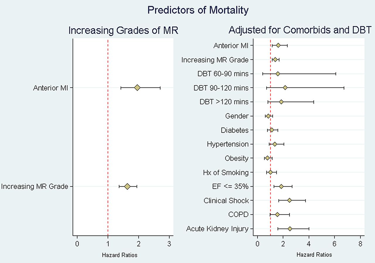
Mean follow-up was 5.9±2.1 years. At 5 years, 40 patients (8%) and 31 patients (10%) were lost to follow-up in the inferior and anterior STEMI groups, respectively. Thirty-day, 1-year and 5-year mortality rates were higher in group 1 (anterior STEMI) compared with group 2 (inferior STEMI; 14.9% vs 6.8%, 26.4% vs 14.3% and 39.7% vs 24.8%, respectively, p<0.001 for all). When the analysis was performed for each grade of IMR between anterior and inferior STEMI, anterior was associated with worse outcomes on every grade of IMR compared with inferior STEMI (figure 1). Kaplan-Meier curves showing long-term survival for the two groups are shown in figure 2. Multivariate Cox survival analysis for predictors of long-term mortality is shown in table 4 and figure 3. After adjusting for age, gender, comorbidities, ejection fraction, grade of IMR and DBT, anterior STEMI was associated with worse outcomes (HR 1.62 (95% CI 1.23 to 2.12), p<0.001).
Multivariate analysis with proportional hazards of 1-year mortality. Predictors of mortality by location of STEMI adjusted for comorbidities and DBT

Thirty-day, 1-year and 5-year mortality rates in anterior versus inferior STEMI across different grades of MR. MR, mitral regurgitation; STEMI, ST elevation myocardial infarction.

Kaplan-Meier curve showing long-term survival in patients with ischaemic MR presenting with anterior STEMI and inferior STEMI. MI, myocardial infarction; MR, mitral regurgitation; STEMI, ST elevation myocardial infarction.

Multivariate Cox survival analysis with predictors of mortality after adjusting for comorbidities and DBT. COPD, chronic obstructive pulmonary disease; DBT, door-to-balloon time; EF, ejection fraction; MI, myocardial infarction; MR, mitral regurgitation; STEMI, ST elevation myocardial infarction.
Discussion
In this study, we demonstrate that overall incidence of IMR by transthoracic 2D echocardiography in patients presenting with acute STEMI who undergo PPCI is 17.8% in anterior STEMI compared with 21.8% in inferior STEMI. We also demonstrate that most common direction of jet in anterior STEMI is central while in inferior STEMI is posterior then central. In addition, IMR in anterior STEMI is associated with worse short-term and long-term outcomes than inferior STEMI across all grades of MR even after adjusting for age, gender, comorbidities, grade of IMR, ejection fraction and DBT.
Higher incidence of IMR in inferior than anterior STEMI was shown in previous studies;8 ,20 ,28 ,32 however, overall numbers in these studies were much higher. This could be explained by the fact that in these studies, many of the patients got thrombolytic therapy or late PCI rather than PPCI, and it has been previously reported that PPCI lowers the incidence of MR in STEMI patients.21 Also, patients with trace MR were not included in our study; had these been included, this might have increased our reported incidence.
It is known that there are two major types of mitral valve apparatus changes that can happen after acute MI, the first is the asymmetric remodelling, where posterior tethering of both mitral leaflets occurs, resulting in posterior bending of the posterior leaflet with over-ride of the anterior leaflet, the resulting IMR jet is usually directed posteriorly. The second is the symmetric remodelling where there is apical tethering of both leaflets with restricted motion of anterior leaflet resulting in apical tenting with apically displaced coaptation line. This results in centrally directed IMR jet. The former occurs mostly in inferior MI while the latter occurs more commonly in anterior MI.34 ,35 This is similar to what was found regarding direction of IMR jet our study.
MI can cause MR by different mechanisms such as changes in LV geometry, mitral valve annulus dilation, LV dysfunction reducing systolic closing pressure, or functional disruption of the posteromedial papillary muscle by displacement increasing tethering forces (tenting). The interplay between these different factors determines the severity of the MR.29–32 ,36 ,37 Inferior MI causes distortion to the mitral valve apparatus more frequently resulting in incompetent valve compared with anterior STEMI. Anterior MI causes MR due to apical tethering of the valve, a mechanism that requires LV dilation. Therefore, MR in anterior STEMI is frequently associated with more dysfunctional LV compared with inferior STEMI. Further, while apical tethering of the leaflets and Mitral valve (MV) annular deformation and dilation are seen in inferior and anterior MI, it has been shown that the severity of these deformities is greater in patients with anterior MI.38 ,39 This is an important distinction as the systolic tenting area of the mitral valve, mitral annular area and spherical index are all independent determinants of regurgitant orifice area.40 Whether it is these differences that result in the difference in outcomes between both types of STEMI is not known, but our novel finding here that IMR has worse outcomes with anterior compared with inferior STEMI may provide a rationale to explore these mechanistic differences in future studies.
Associations of increased 30-day mortality10 and 1-year mortality1 ,3–5 ,10 ,23 with IMR have been previously reported. Comparisons were carried out between two groups (non-significant vs significant IMR) or three groups (no MR, mild MR and moderate/severe MR). Our study is the first to compare between IMR regarding index STEMI location. This might be because of the large sample size of our study which provided enough power to perform this comparison. Outcomes of anterior STEMI overall is known to be worse than inferior STEMI, but in our study, there was no difference between both groups in all risk factors, clinical presentation, comorbidities and coronary intervention. Even after adjusting for known factors associated with more mortality, still IMR in anterior STEMI showed worse prognosis.
To the best of our knowledge, our study is the first to date to compare outcomes of IMR by STEMI location in a large population of patients >800 patients who underwent PPCI within the first 12 hours of presentation and had a mean follow-up of 5.9 years.
Study limitations
As with any retrospective, single-centre analysis, our study may be limited by selection bias and the results might not be generalisable to other centres or hospitals. A second limitation is that our patient population spans a 20-year period, over which advances in coronary intervention and cardiac care were tremendous. A third limitation is that we did not have grades of MR in follow-up echocardiography, as several studies before reported that the severity of MR changes over time in the subacute phase of the MI.17 ,24 In addition, we did not have data on clinical follow-up such as heart failure readmissions, or number of patients who received surgical intervention for the MR in follow-up, and this might have affected noticed outcomes. Another limitation is that patients who presented with STEMI and died before performing an echocardiogram were not accounted for and the presence of IMR in these patients was not known.
Conclusion
IMR occurs more commonly with inferior compared with anterior STEMI. However, IMR with anterior had much worse outcomes in every grade of regurgitation compared with inferior STEMI.
References
Footnotes
Competing interests None declared.
Ethics approval Institutional Review Board (IRB) of Cleveland Clinic.
Provenance and peer review Not commissioned; externally peer reviewed.
Data sharing statement No additional data are available.