Article Text

Abstract
Objective The prognosis of apical hypertrophic cardiomyopathy (APH) has been benign, but apical myocardial injury has prognostic importance. We studied functional, morphological and electrocardiographical abnormalities in patients with APH and with apical aneurysm and sought to find parameters that relate to apical myocardial injury.
Methods Study design: a multicentre trans-sectional study. Patients: 45 patients with APH and 5 with apical aneurysm diagnosed with transthoracic echocardiography (TTE) in the database of Hamamatsu Circulation Forum. Measure: the apical contraction with cine-cardiac MR (CMR), the myocardial fibrotic scar with late gadolinium enhancement (LGE)-CMR, and QRS fragmentation (fQRS) defined when two ECG-leads exhibited RSR’s patterns.
Results Cine-CMR revealed 27 patients with normal, 12 with hypokinetic and 11 with dyskinetic apical contraction. TTE misdiagnosed 11 (48%) patients with hypokinetic and dyskinetic contraction as those with normal contraction. Apical LGE was apparent in 10 (83%) and 11 (100%) patients with hypokinetic and dyskinetic contraction, whereas only in 11 patients (41%) with normal contraction (p<0.01). Patients with dyskinetic apical contraction had the lowest left ventricular ejection fraction, the highest prevalence of ventricular tachycardia, and the smallest ST depression and depth of negative T waves. The presence of fQRS was associated with impaired apical contraction and apical LGE (OR=8.32 and 8.61, p<0.05).
Conclusions CMR is superior to TTE for analysing abnormalities of the apex in patients with APH and with apical aneurysm. The presence of fQRS can be a promising parameter for the early detection of apical myocardial injury.
This is an Open Access article distributed in accordance with the Creative Commons Attribution Non Commercial (CC BY-NC 4.0) license, which permits others to distribute, remix, adapt, build upon this work non-commercially, and license their derivative works on different terms, provided the original work is properly cited and the use is non-commercial. See: http://creativecommons.org/licenses/by-nc/4.0/
Statistics from Altmetric.com
Key messages
-
What is already known about this subject?
-
Apical injury in hypertrophic cardiomyopathy (HCM) has poor outcome. Cardiac MRI can detect regional myocardial hypertrophy not readily recognised with echocardiography in patients with HCM.
-
What does this study add?
-
Cardiac MRI is superior to echocardiography for detection of functional and morphological abnormalities of the left ventricular apex in patients with apical hypertrophy and with apical aneurysm. The presence of fragmented QRS can help to detect apical myocardial injury.
-
How might this impact on clinical practice?
-
To precisely estimate apical morphology and function and for early detection of apical myocardial injury in HCM.
-
To apply fragmented QRS as an early indicator of apical injury in HCM.
Introduction
Apical hypertrophic cardiomyopathy (APH) is an uncommon phenotype of hypertrophic cardiomyopathy (HCM).1 ,2 The incidence of APH is only 1–2% of HCM in Western countries, but is reported to be up to 25% in Japan.1 ,3
The prognosis of APH seems to be benign, but apical myocardial injury such as apical aneurysm and fibrosis has been related to adverse cardiac events.3–5 Although the detailed mechanisms for the development of apical aneurysm remain unknown, it is crucial to precisely analyse the functional and histological features of the left ventricular (LV) apex in patients with HCM.
Two-dimensional transthoracic echocardiography (TTE) is still the standard for the diagnosis of HCM, but TTE has limitations for evaluating morphology and function of the LV apex. Cardiac MRI (CMR) is now established to assess cardiac function with a high spatial resolution of cine-CMR, and to differentiate fibrosis from normal myocardium with late gadolinium enhancement (LGE)-CMR.6 Many studies have elucidated the diagnostic and prognostic values of CMR in HCM,7–12 but few reports showed the clinical relevance in APH and apical aneurysm.4 ,7 ,13
Since CMR is not necessarily available in all institutes and for all patients, and has a problem of cost, it is also necessary to predict apical myocardial injury with other imaging modalities including a 12-lead ECG. Giant negative T waves (GNT) with ST segment depression have been recognised as an index of apical myocardial injury and APH.1 ,2 ,14 ,15 Several recent studies have suggested that the region of a myocardial scar is associated with a fragmentation of QRS complexes (fQRS) that can be a marker of a prior myocardial infarction with a substantially higher sensitivity compared with the Q waves.16 ,17
For the precise estimation of apical morphology and function and for early detection of apical myocardial injury in HCM, we investigated apical contraction and LGE in patients with APH and with apical aneurysm using CMR, and examined the association with clinical, TTE and ECG features.
Patients and methods
Patients
This was a multicentre trans-sectional study, and comprised 48 patients consecutively with APH and 5 with apical aneurysm who underwent CMR and were registered to the database of Hamamatsu Circulation Forum between 2004 and 2012. Patients were initially suspected to have APH because of typical ECG abnormalities with or without LV hypertrophy in other LV segments. Apical morphologies were basically diagnosed using TTE by a cardiologist or cardiac sonographer in each institute. APH was determined when apical wall thickness >15 mm, or ratio of apical/basal wall thickness >1.3.13 Apical aneurysm was defined as a discrete thin-walled dyskinetic or akinetic segment of the most distal portion of the LV chamber.4 ,5
Patients with uncontrollable hypertension and with severe valvular diseases were not registered to the database. Obstructive atherosclerotic coronary artery disease was excluded by the absence of significant coronary arterial narrowing (>50% stenosis) at coronary cine or computed tomographic angiography (n=14 for APH and n=4 for apical aneurysm), or in the remaining patients, by absent histories of chest pain, coronary risk factors and acute coronary syndrome. All patients underwent CMR within 1 month after diagnosis with TTE.
Three patients with complete bundle branch blocks (BBBs) were then excluded from the study in consideration of modification of apical wall motion. Finally, this study consisted of 50 patients (45 patients with APH and 5 with apical aneurysm), and the database was analysed retrospectively. This study protocol was conducted in accordance with the Declaration of Helsinki.
Protocol for CMR
CMR was performed on 1.5 tesla (T) MR systems (Enshu Hospital: Excelart Vantage 1.5 T, Toshiba, Japan; Hamamatsu University Hospital: Signa Infinity Twinspeed, GE Medical Systems, Waukesha, USA; Hamamatsu Red Cross Hospital: Acheiva 1.5 T, Phillips Inc, Bothell, USA; Kosai General Hospital: Vantage Titan, Toshiba, Japan; and Seirei Mikatahara General Hospital: Signa Horizon Release 5.7, GE Medical Systems). The details of MR systems are described elsewhere.10 ,18 Typically, two-dimensional (2D) FIESTA and LGE images were acquired in the short axis, vertical long axis and horizontal long axis orientations. The slice thickness/gap was typically 10 mm/0 mm (6–9 slices).
Breath-hold cine MRIs were obtained in contiguous short-axis planes from apex to base of the heart with the patient in a resting state. The 2D FIESTA cine images were based on the steady state free precession sequence. The imaging parameters were as follows: matrix of 192×192, field of view of 34 cm, flip angle of 45° and readout bandwidth of 125 kHz. Sixteen data lines were acquired per segment.
LGE images were acquired 15 min after an injection of 0.2 mmol/kg of contrast material (Gd-DTPA-BMA, Fuji Pharma, Tokyo, Japan). LGE imaging was based on the inversion recovery prepared fast gradient echo (IR-FGRE) sequence. The imaging parameters were as follows: matrix of 256×160, field of view of 34 cm, flip angle of 20° and readout bandwidth of 31.25 kHz. The IR-FGRE technique was repeated during every R-to-R interval and the trigger delay was 300 ms. The readout data line was 160 each, where 24 data lines were acquired per segment. The optimum inversion time (200–240 ms) was measured right before the LGE imaging.
Analyses of CMR
Two experienced cardiovascular radiologists (KS and HS) interpreted all the CMRs without knowledge of clinical findings. LV end-diastolic volume (LVEDV), end-systolic volume (LVESV), left ventricular ejection fraction (LVEF) and LV mass (LVM) were acquired from the 2D FIESTA cine images in short axis view. The values for LV volume and mass were indexed by dividing them with body surface area (LVEDV index, LVESV index and LVM index).
We classified patients into three groups according to apical contraction: (1) normal: apical hypertrophy and spade-like morphology at end-diastole and a disappearance of apical cavity at end-systole, (2) hypokinetic: apical hypertrophy and spade-like morphology at end-diastole with retention of apical cavity at end-systole and (3) dyskinetic: apical wall thinning and dyskinetic apical contraction (with or without ventricular obstruction; figures 1⇓–3). Regional analyses of LGE-CMR were performed using the 17-segment model.15 Then we examined the presence of LGE in each segment, determined the presence or absence of apical LGE and counted the number of segments with LGE. The apical injury was defined to be impaired apical contraction (hypokinetic or dyskinetic contraction) or the presence of apical LGE. The apical contraction and the presence, location and extent of LGE were determined by the consensus of the two observers with analyses of all the orientations.

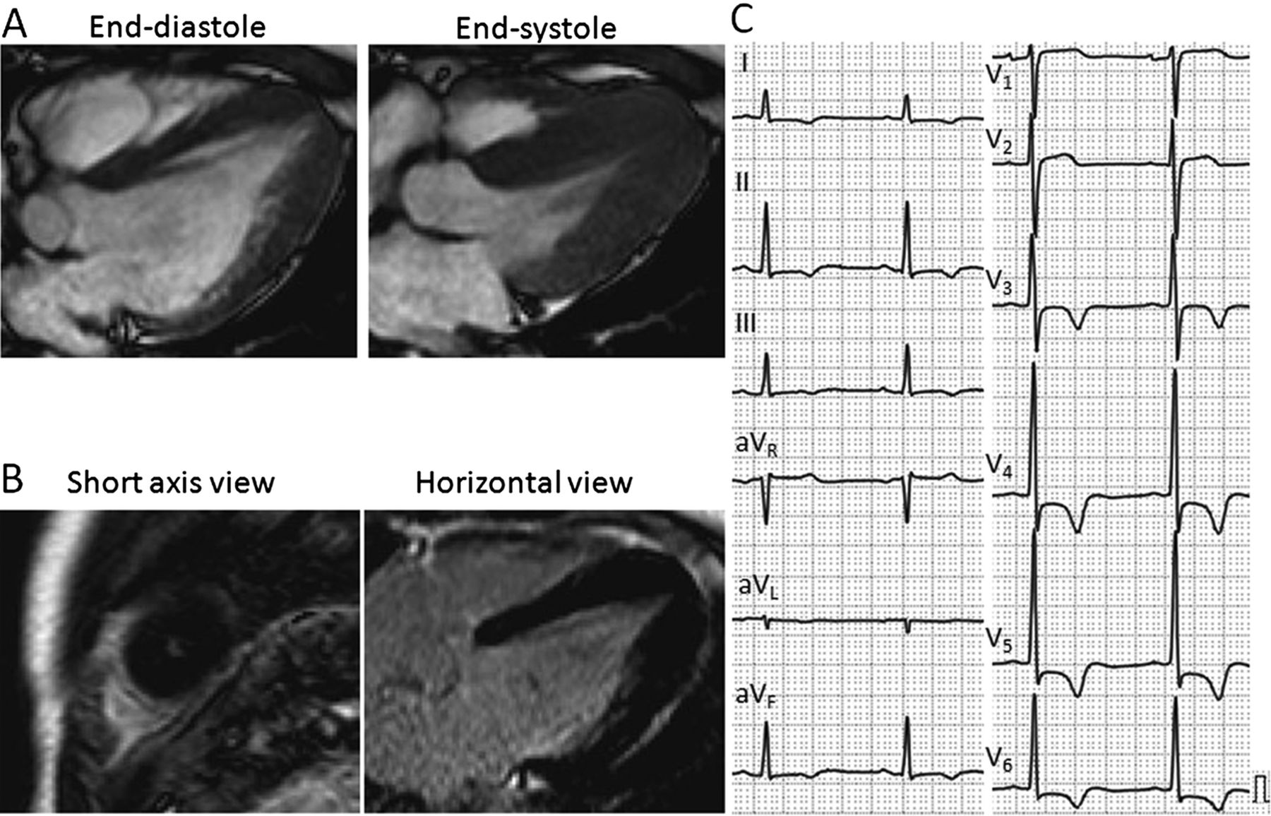
A representative patient with normal apical contraction. (A) Cine-cardiac MRIs (CMRs) at end-diastole (left) and end-systole (right). Apical hypertrophy and spade-like morphology at end-diastole and a complete disappearance of apical cavity at end-systole were shown. (B) Late gadolinium enhancement (LGE)-CMRs at short-axis view (left) and horizontal long-axis view (right). No LGE was observed. (C) Standard 12-lead ECG. High voltage QRS complexes, strain type ST depression and giant negative T waves were apparent.
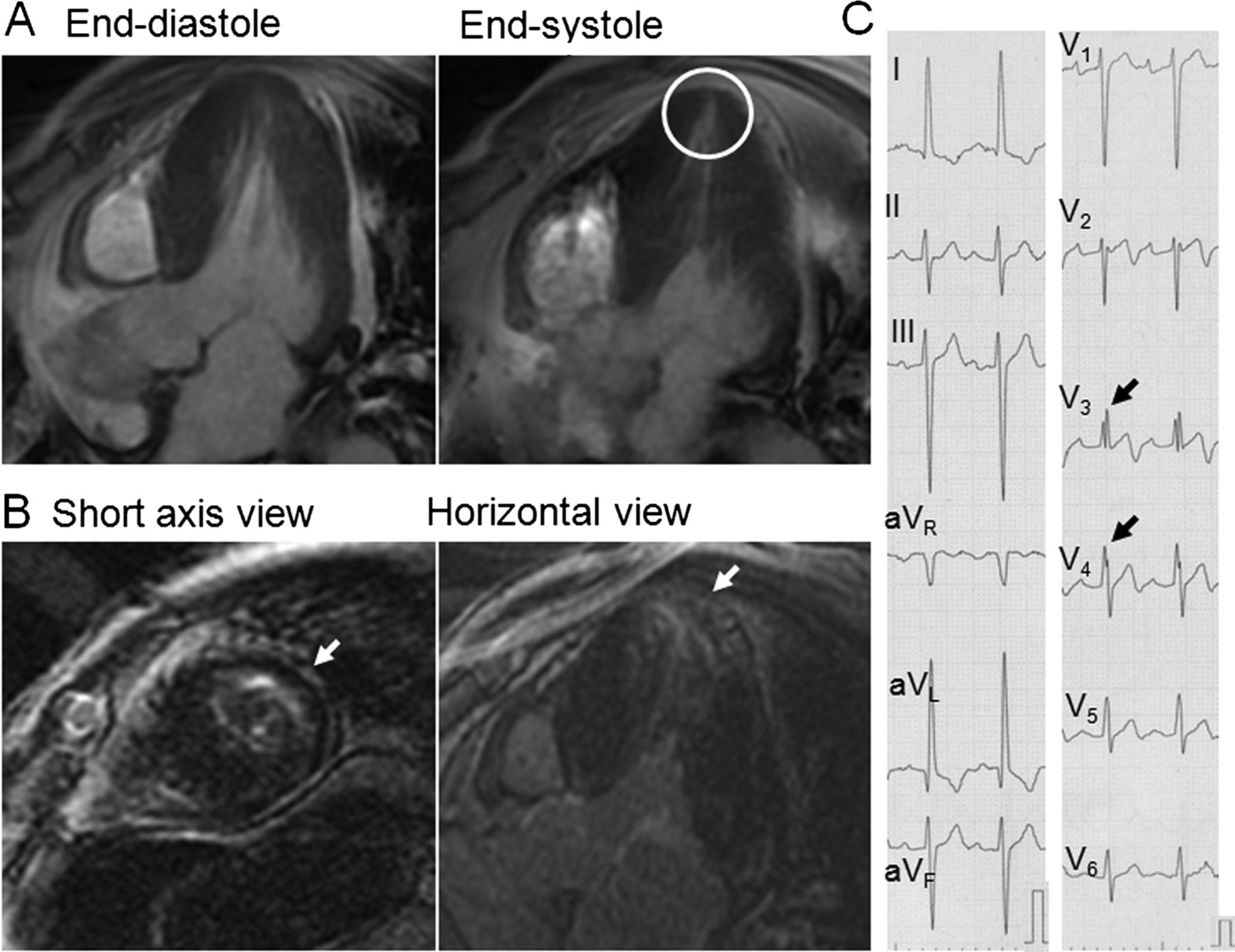
A representative patient with hypokinetic apical contraction. (A) Cine-cardiac MRIs (CMRs) at end-diastole (left) and end-systole (right). Apical hypertrophy and spade-like morphology at end-diastole were similar to those in a patient with normal apical contraction, but the apical cavity retained and did not completely disappear at end-systole (white circle). (B) Late gadolinium enhancement (LGE)-CMRs at short-axis view (left) and horizontal long-axis view (right). LGE at the left ventricular apex was apparent (white arrows). (C) Standard 12-lead ECG. In addition to giant negative T waves, fragmented QRS complexes at V3 and V4 leads were apparent (black arrows).

A representative patient with dyskinetic apical contraction. (A) Cine-cardiac MRIs (CMRs) at end-diastole (left) and end-systole (right). The apical wall thinning, dyskinetic apical contraction and apical pouch were apparent. (B) Late gadolinium enhancement (LGE)-CMR images at short-axis view (left) and horizontal long axis view (right). LGE at the left ventricular apex was apparent (white arrows). (C) Standard 12-lead ECG. In addition to high voltage QRS complexes and giant negative T waves, fragmented QRS complexes at III, aVL and aVF leads were apparent (black arrows).
Analyses of 12-lead ECG and TTE
Twelve-lead ECG and TTE were performed before CMR (<1 month) in all patients. We examined the presence of fQRS, which was defined when notched R or S were present in at least two-ECG leads (figure 2 and 3).16 The apical motion was analysed with 2D mode of TTE (apical 2 and 4 chamber views) by a cardiologist in each institute. Patients also underwent a 24 h ambulatory Holter ECG. The intervals between the analyses by CMR and Holter ECG were <3 months. Non-sustained ventricular tachycardia (VT) and supraventricular tachycardia (SVT) were defined as three or more consecutive premature complexes with a heart rate of >100 bpm.
Statistical analysis
All data were expressed as the means±SD of the indicated numbers (n) or percentages, as appropriate. Categorical variables were compared among the groups by χ2 analysis. Continuous variables were compared by unpaired t test or one-way analysis of variance followed by Scheffe's post hoc analysis. Differences were considered to be significant when p<0.05. In the multivariate analysis, only ECG or TTE parameters that showed p<0.1 in the univariate analyses were incorporated. The sensitivity and specificity of fQRS for impaired apical contraction and apical LGE were derived from dividing the patient numbers with or without fQRS from those with or without impaired apical contraction or apical LGE. All the statistical analyses were performed using software StatFlex (V.6).
Results
Estimation of apical contraction
All patients were assigned to three groups according to apical contraction estimated with cine-CMR: normal (n=27), hypokinetic (n=12) and dyskinetic (n=11) apical contraction, respectively. TTE underestimated 6 (50%) and 5 (45%) patients with hypokinetic and dyskinetic apical contraction in cine-CMR as those with normal apical contraction, and also 1 (9%) patient with dyskinetic apical contraction as hypokinetic apical contraction. Additionally, TTE overestimated 2 (7%) patients with normal apical contraction as those with hypokinetic apical contraction, and 1 (8%) patient with hypokinetic apical contraction as dyskinetic apical contraction (see online supplementary table S1). There was no tendency in the prevalence of misdiagnosis among the institutes or analysts. Apical thrombus was detected in one patient with dyskinetic apical contraction by TTE and CMR.
Representative images
Figures 1⇑–3 show representative cine-CMRs and LGE-CMRs, and 12-lead ECGs in patients with normal, hypokinetic and dyskinetic apical contraction. LGE-CMRs exhibited LGE at the LV apex in patients with hypokinetic and with dyskinetic apical contraction, but did not in the patient with normal apical contraction. The 12-lead ECGs demonstrated high voltage QRS complexes, strain type ST depression and GNT in the patients, although the degree of ST depression and the depth of GNT were to a lesser extent in patients with hypokinetic and with dyskinetic apical contraction. Contrarily, fQRS was present in only patients with hypokinetic and with dyskinetic apical contraction.
CMR findings
Table 1 demonstrates the differences in CMR parameters among the three patient groups. LVEF was significantly lower in patients with dyskinetic apical contraction than those with normal or hypokinetic apical contraction, whereas LV volume, mass or maximum segmental wall thickness did not differ. LGE in the LV apex was found in all patient groups, but the prevalence was relatively low in patients with normal apical contraction (41%), whereas most patients with hypokinetic apical contraction (83%) and all patients with dyskinetic apical contraction (100%) had LGE in the LV apex. There was no difference in the number of LGE segments among the groups.
CMR parameters in patients with normal, hypokinetic and dyskinetic apical contraction
Clinical features
We then compared clinical features, TTE and ECG parameters among patients with different apical contraction and between patients with and without apical LGE (tables 2⇓–4 and see online supplementary tables S2–4). The prevalence of VT and medication with β-blockers were highest in patients with dyskinetic apical contraction. The prevalence of VT, SVT, and medication with antiarrhythmic agents and antithrombotic agents was higher in patients with apical LGE. No patients had signs of systemic thromboembolism. Age, sex, symptoms, family history and serum NT-proBNP level did not significantly differ.
Comparison of clinical features in terms of apical contraction
Comparison of TTE parameters in terms of apical contraction
Comparison of ECG parameters in terms of apical contraction
TTE parameters did not significantly differ among patients with different apical contraction or between patients with and without apical LGE. In 12-lead ECG, the maximum ST depression and maximum depth of GNT were smaller in patients with dyskinetic apical contraction than those in the other two groups. The prevalence of fQRS was higher in patients with hypokinetic or with dyskinetic apical contraction than that in patients with normal apical contraction. The smaller maximum ST depression and higher prevalence of fQRS were also found in patients with apical LGE. fQRS was present dominantly at inferior leads (III, aVF) and mid-precordial leads (V3, V4; see online supplementary table S5). Patients with fQRS had a higher prevalence of VT than those without fQRS (38% vs 14%, p<0.05).
Multivariate analyses for apical contraction and apical LGE
The multivariate analyses showed that fQRS was the sole index of impaired apical contraction (hypokinetic and dyskinetic apical contraction) and apical LGE (table 5), but was not an index of dyskinetic apical contraction (data not shown). The sensitivity and specificity of fQRS were 65.2% (15/23 patients) and 77.8% (21/27 patients) for impaired apical contraction, and 59.4% (19/32 patients) and 88.9% (16/18 patients) for apical LGE, respectively. The PR interval was also an index of impaired apical contraction, although it did not reach significance in the univariate analysis.
Multivariate analyses in terms of apical contraction and apical LGE
Discussion
We examined apical morphology and function in patients with APH and with apical aneurysm and showed that: (1) cine-CMR could identify impaired apical contraction more precisely compared with TTE, (2) the prevalence of apical LGE was higher in patients with hypokinetic and dyskinetic apical contraction than in those with normal apical contraction, and (3) the presence of fQRS was associated with impaired apical contraction and apical LGE. We could emphasise the clinical usefulness of CMR for estimating apical morphology and function, and provide, for the first time, the relevance of fQRS for prediction of apical myocardial injury.
APH and apical aneurysm in HCM
APH has a benign prognosis in Japan, with the exception of elderly patients, but the condition appears to be less benign in Western countries.3 ,5 ,19 The prognosis of patients with HCM becomes quite poor when the apex contains aneurysms. In a report from Maron et al,4 43% of patients with apical aneurysm experienced adverse cardiac events. The intraventricular pressure gradient due to mid-ventricular obstruction can trigger chronic myocardial ischaemia, which in turn results in apical aneurysm formation.20 The severe apical hypertrophy could also impose pressure overload and coronary flow impairment of the LV apex, which may thereby provoke myocardial infarction and/or fibrosis.21 All patients with dyskinetic apical contraction had apical LGE, although there was no difference in the thickness of interventricular septum or posterior wall. Patients with dyskinetic apical contraction and those with apical LGE had higher prevalence of VT. Thus, myocardial fibrosis at the LV apex was closely associated with dyskinetic apical contraction and development of VT.
Estimation of apical abnormalities with TTE and CMR
Although TTE has been the standard tool for the diagnosis of HCM, it has limitations for precise visualisation of whole ventricles and quantification of hypertrophy, especially for the LV apex. In addition to apical views, a careful approach with sequential parasternal short axis view is required. CMR can image in any plane and cine-CMR has achieved high spatial resolution with a substantial improvement of blood to myocardium contrast. Since Moon et al7 reported a series of patients in whom CMR could detect unrecognised APH, several studies have showed the usefulness of CMR for detection of APH and apical aneurysm.4 ,13 ,15 ,22 We compared apical contraction with TTE and cine-CMR in patients who were recognised as having APH or apical aneurysm. In addition to dyskinetic apical contraction, we defined hypokinetic apical contraction when the apical cavity retained at end-systole. This estimation was quite novel, and TTE underestimated 50% and 45% patients with hypokinetic and dyskinetic apical contraction, respectively, as normal. The misdiagnosis was not ascribed to different institutes or analysts. Maron et al4 studied 28 of 1299 patients with HCM who had apical aneurysm, and TTE could identify apical aneurysm in only 16 patients, but CMR could detect it in the remaining 12 patients.
The high prevalence and particular distribution patterns of LGE in the LV wall further emphasise the clinical usefulness of CMR for the diagnosis of HCM.10 ,15 ,23 ,24 Although apical aneurysms relate to adverse cardiac events, the clinical implications of hypokinetic apical contraction in APH remain undefined. In this study, most patients with hypokinetic apical contraction had LGE in the LV apex. The higher prevalence of LGE in the LV apex in patients with hypokinetic apical contraction suggests more advanced myocardial fibrosis than in those with normal apical contraction. Although there is no evidence that patients with hypokinetic apical contraction progressively develop apical aneurysm and/or adverse cardiac events, early detection of apical myocardial injury with CMR may help for risk stratification and management of APH. Patients with hypokinetic apical contraction, but without LGE, may benefit from aggressive therapies with β-adrenergic receptor blockers or inhibitors of renin-angiotensin-aldosterone systems.
There were no differences in standard TTE parameters in terms of apical contraction and apical LGE, although the mid-ventricular hypertrophy was expected to have caused a chronic intraventricular pressure gradient and, as a result, apical myocardial injury. The reason for the lack of differences remains unknown, but may be dependent on heterogeneous LV hypertrophy in such patients.22
Prediction of apical myocardial injury with 12-lead ECG
In HCM, a 12-lead ECG shows a wide variety of abnormal patterns and provides diagnostic and prognostic information. We, as well as Dumont et al15 ,25 have exhibited correlation between LGE and conduction disturbance, abnormal Q waves and GNT. However, studies for ECG abnormalities that relate to apical myocardial injury remain inadequate.
GNT are recognised as the most typical feature of APH, at least in Japanese patients.1 ,2 The size of GNT has been related to the apical wall thickness and asymmetric distal hypertrophy.14 ,15 The maximum ST depression was smaller in patients with dyskinetic apical contraction and with apical LGE than those in other groups. The maximum depth of GNT was smaller in patients with dyskinetic apical contraction, but did not differ between patients with and without apical LGE. Previous studies showed a disappearance of GNT during a long-term follow-up.26 ,27 Given that the relative myocardial ischaemia caused ST-T abnormalities in patients with APH,21 ,28 the apical wall thinning and fibrosis can be mechanisms of the decreases in ST depression. However, the correlation between the depth of GNT and apical fibrosis remains controversial.15 ,29 The reason why PR interval became significant in the multivariate analysis for impaired apical contraction is also uncertain.
However, the most important finding was that fQRS, without an evidence of BBB, was associated with impaired apical contraction and apical LGE. Actually, fQRS at inferior and/or mid-precordial leads was apparent in several previous case reports showing apical aneurysm formation in APH.13 ,20 ,30 The finding that fQRS was also related with hypokinetic apical contraction suggested that fQRS could be an index of myocardial injury before the formation of apical aneurysm. Various prior studies have suggested that the region of a myocardial scar is associated with alteration in QRS morphology, leading to a terminal conduction delay or a fragmentation of QRS complexes.16 ,17 Das et al16 showed that fQRS is a marker of a prior myocardial infarction, defined by regional perfusion abnormalities, that has a substantially higher sensitivity and negative predictive value compared with the Q waves. We showed moderately high sensitivity and specificity of fQRS for detection of apical myocardial injury. Das et al16 also reported that the fQRS is an independent predictor of cardiac events in patients with coronary arterial disease. In this study, fQRS was associated with a higher prevalence of VT. Despite the analysis in small patient groups and the lack of prognostic information, fQRS can be a promising parameter for the early detection of apical myocardial injury and the start of treatment in patients with APH.
Limitations
This study consisted of patients who underwent CMR at five different institutes. The selection bias, different equipment and small sample size may be limitations for extrapolating our data to diverse patient groups. Coronary angiography was not routinely performed to exclude coronary arterial disease, but a section of patients were diagnosed by a lack of symptoms or risk factors. Although there were no significant stenotic coronary lesions in patients with apical aneurysm, the possibility of coronary spasm or coronary embolism cannot be excluded. We evaluated apical contraction and apical LGE as indices of apical injury. The estimation of apical wall thickening and the quantification of apical fibrosis (possibly with T1 mapping) may provide more precise information. Finally, we did not examine the relevance of apical myocardial injury in terms of adverse cardiac events during a long-term follow-up.
Conclusions
CMR was superior to TTE for detection of functional and morphological abnormalities of the LV apex in patients with APH and with apical aneurysm. The presence of fQRS can help for suspected apical myocardial injury. More advanced diagnostic and prognostic evaluation in a larger population can clarify the clinical relevance of CMR and fQRS in those patients.
References
Supplementary materials
Supplementary Data
This web only file has been produced by the BMJ Publishing Group from an electronic file supplied by the author(s) and has not been edited for content.
Files in this Data Supplement:
- Data supplement 1 - Online tables
Footnotes
-
Contributors KS was involved in description of manuscript. HS was involved in description and revision of manuscript. MS and MS were involved in analysis of MRI data. MN and TS were involved in analysis of echo and ECG data. TU, HK, HO, YW, KT, HT and HT were involved in enrolment of patients. YT and HS were involved in MRI technical support. HH was involved in general control of the study.
-
Funding This research received no specific grant from any funding agency in the public, commercial or not-for-profit sectors.
-
Competing interests None.
-
Patient consent Obtained.
-
Ethics approval Hamamatsu Univ Sch Med.
-
Provenance and peer review Not commissioned; externally peer reviewed.
-
Data sharing statement No additional data are available.