Article Text

Abstract
Background: Factors associated with impaired clinical status in a cross-sectional study of patients with repaired tetralogy of Fallot (TOF) have been reported previously.
Objectives: To determine independent predictors of major adverse clinical outcomes late after TOF repair in the same cohort during follow-up evaluated by cardiac magnetic resonance (CMR).
Methods: Clinical status at latest follow-up was ascertained in 88 patients (median time from TOF repair to baseline evaluation 20.7 years; median follow-up from baseline evaluation to most recent follow-up 4.2 years). Major adverse outcomes included (a) death; (b) sustained ventricular tachycardia; and (c) increase in NYHA class to grade III or IV.
Results: 22 major adverse outcomes occurred in 18 patients (20.5%): death in 4, sustained ventricular tachycardia in 8, and increase in NYHA class in 10. Multivariate analysis identified right ventricular (RV) end-diastolic volume Z ⩾7 (odds ratio (OR) = 4.55, 95% confidence interval (CI) 1.10 to 18.8, p = 0.037) and left ventricular (LV) ejection fraction <55% (OR = 8.05, 95% CI 2.14 to 30.2, p = 0.002) as independent predictors of outcome with an area under the receiver operator characteristic curve of 0.850. LV ejection fraction could be replaced by RV ejection fraction <45% in the multivariate model. QRS duration ⩾180 ms also predicted major adverse events but correlated with RV size.
Conclusions: In this cohort, severe RV dilatation and either LV or RV dysfunction assessed by CMR predicted major adverse clinical events. This information may guide risk stratification and therapeutic interventions.
Statistics from Altmetric.com
Surgical management of tetralogy of Fallot (TOF) results in residual haemodynamic abnormalities in most patients. Relief of the right ventricular (RV) outflow tract obstruction induces pulmonary regurgitation (PR) and chronic volume loading that leads to progressive RV dilatation, dysfunction and symptoms.1–6 Although the haemodynamic burden from chronic RV volume load can be tolerated without symptoms during childhood, a growing body of evidence suggests that the incidence of ventricular dysfunction, arrhythmia, heart failure and death increases substantially in adult survivors of TOF repair.3 7 Recent studies on large cohorts who had undergone TOF repair and had been followed up for >30 years demonstrated that during the third postoperative decade the annualised risk of death more than tripled, increasing from 0.27% to 0.94%.8 9 Although pulmonary valve replacement (PVR) has been proposed as a treatment for symptomatic patients, several investigators noted that placing a pulmonary valve in patients with severe RV dilatation and dysfunction may not lead to adequate functional recovery of the RV.7 10 11 These observations have led several centres to advocate PVR before the onset of symptoms to preserve RV mechanics.7 12 13 Despite greater awareness of the natural history of patients with repaired TOF, there continues to be uncertainty about the optimal timing of re-intervention. Specifically, there is a paucity of information on predictors of adverse clinical outcomes in these patients. Such information may provide the rationale for intervention in patients at high risk.
We previously published the results of a cross-sectional study evaluating factors associated with adverse clinical status in 100 consecutive long-term survivors of TOF repair evaluated by cardiac magnetic resonance (CMR).4 The goal of the present study was to identify independent predictors of major adverse clinical outcomes in this cohort 3–7 years after baseline evaluation.
SUBJECTS AND METHODS
Subjects
The study was designed as a single-centre cohort investigation. Patients enrolled in the previous cross-sectional study between 1 November 1997 and 31 August 2001 fulfilled the following criteria: (a) repaired TOF with pulmonary stenosis or atresia; (b) ⩾10 years between complete repair and baseline evaluation; (c) completed standardised CMR examination protocol; and (d) concurrent clinical evaluation.4 Of these 100 patients evaluated at baseline, 88 had ⩾2 months’ follow-up (median 4.2 years, range 2 months–7.3 years) and comprised the present study group. For patients who underwent PVR during the study period, follow-up was censored at the time of the most recent preoperative evaluation. Of the 12 patients with <2 months’ follow-up, three underwent PVR immediately after baseline evaluation and nine were lost to follow-up. The Children’s Hospital Committee on Clinical Investigations approved review of the medical records and computer databases.
Clinical data
The following demographic and clinical data were abstracted from patients’ medical records: date of birth, gender, date and age at each surgical procedure, type of surgical procedures (categorised as shunt, transannular patch repair, non-transannular patch repair, and right ventricle-to-pulmonary artery conduit repair), age at CMR, and time from TOF repair to baseline evaluation. The following variables were recorded from baseline and most recent clinical evaluations: height and weight, vital signs, symptoms (eg, shortness of breath, exertional dyspnoea, palpitations, syncope, chest pain), New York Heart Association (NYHA) class, findings on physical examination (eg, tachypnoea, gallop rhythm, jugular venous distention, hepatomegaly, peripheral oedema), and medications (categorised as digoxin, diuretics, angiotensin converting enzyme inhibitors, and antiarrhythmic agents). In addition, the presence of comorbidities such as lung, liver, or renal abnormalities was noted.
Cardiac magnetic resonance
The CMR protocol used in this study was previously described in detail.4 Briefly, studies were performed with a commercially available 1.5 T scanner (Signa Horizon; GE Medical Systems, Milwaukee, WI, USA). Biventricular volumes and function were assessed using a breath-hold, ECG-triggered, segmented k-space, fast spoiled gradient recalled cine sequence in two- and four-chamber planes followed by 12 contiguous short-axis slabs perpendicular to the long axis of the left ventricle extending from the plane of the atrioventricular valve through the apex (slice thickness 6–8 mm, interslice space 0–2 mm). PR volume and fraction were assessed by measuring antegrade and retrograde flow in the main pulmonary artery using a free-breathing, ECG-triggered, cine phase velocity pulse sequence.
Left and right ventricular end-diastolic (maximal) and end-systolic (minimal) volumes, mass, stroke volumes, and ejection fraction were measured using commercially available software (MASS; Medis, Leiden, Netherlands) as described by Lorenz.14 Quantification of flow rates and calculation of PR was performed using customised software as previously described.15 RV and left ventricular (LV) end-diastolic volumes and mass were adjusted to body surface area and Z-scores were calculated based on published normal values derived using the same CMR technique.16 In the absence of normal reference data for end-systolic volume, this variable was adjusted to body surface area.
Echocardiography
Echocardiograms at baseline evaluation were reviewed for the presence and severity of tricuspid regurgitation (graded by width of the vena contracta as none or trivial, mild, moderate, or severe).
Rhythm abnormalities
Arrhythmia history was abstracted from patient records, electrophysiology studies and Holter results. A 15-lead electrocardiogram was reviewed at the time of baseline evaluation for rhythm, conduction abnormalities and duration of the QRS complex.
Outcomes
The primary outcome was one or more of the following major adverse clinical events: (a) death; (b) sustained ventricular tachycardia (VT) lasting >30 seconds or requiring cardioversion; (c) increase in NYHA to class III or IV. Secondary outcomes included PVR or placement of an implantable defibrillator, or both. In addition to predictors of these outcomes, we sought to identify predictors of “no clinical progression”, defined as NYHA class I at baseline and follow-up, and no new symptoms or medications at follow-up.
Statistics
Demographic, clinical and laboratory variables were compared for patients who achieved the primary outcome and those who did not using the Fisher exact test for categorical variables and either the Wilcoxon rank sum test or two-sample t test for continuous variables. Multivariate analyses adjusting for length of follow-up since baseline CMR was performed using logistic regression analysis; odds ratios (ORs), 95% confidence intervals (CIs), and areas under the receiver operator characteristic (ROC) curves were calculated for each model. ROC curves were used to identify which cut-off points provided the best combinations of sensitivity and specificity for variables identified by multivariate analysis as being independently associated with the primary outcome. Clinical characteristics at baseline and follow-up were compared using the paired t test for continuous variables and the McNemar test for dichotomous variables. Commercially available statistical software was used for data analysis (STATA version 9.0, College Station, Texas, USA).
RESULTS
Demographic, clinical and laboratory data
Table 1 summarises demographic, clinical and laboratory data of the 88 cohort patients. During a median follow-up of 4.2 years, 24 patients (27%) had worsening NYHA class, 28 patients (32%) had addition of 41 cardiac drugs, 42 patients (48%) had at least one new symptom related to the cardiovascular system, and 37 patients (42%) had new physical examination findings related to the cardiovascular system. In addition, 17 patients (19%) had new-onset atrial flutter or fibrillation or non-sustained VT. Comorbidities were common, including lung disease in 16 (18%), liver disease in 7 (8%), renal disease in 3 (3%), and other chronic non-cardiac conditions in 34 patients (39%).
During follow-up in the 88 patients, 27 patients underwent PVR or conduit revision, 3 had additional cardiac surgical procedures, 24 underwent 29 transcatheter interventions, and 20 had placement of an implantable defibrillator.
Major adverse clinical outcomes
During follow-up, 22 major adverse clinical outcomes were recorded in 18 patients (20.5%). There were four deaths (4.5%); all died suddenly out of hospital. Sustained VT was documented in eight patients (9%), one of whom died suddenly. NYHA class changed from good (I or II) to poor (III or IV) in eight patients and from class III to IV in two additional patients (11%). Among the 10 patients with worsening NYHA class, two died suddenly and one had sustained VT.
Predictors of major adverse clinical outcomes
Table 2 summarises the odds ratios and areas under the ROC curve for the strongest predictors of major adverse clinical outcomes. Gender, anatomic type of TOF (pulmonary stenosis vs atresia), type of repair (RV outflow tract patch vs conduit), and degree of tricuspid regurgitation were not predictive of the primary outcome. Among the CMR variables, PR volume or fraction, RV mass and mass-to-volume ratio, LV end-diastolic volume, LV end-systolic volume, LV mass, and the ratio of RV-to-LV diastolic volume did not predict the primary outcome. Except for jugular venous distention, none of the findings on clinical examination were predictive of the primary outcome, nor were any of the symptoms or comorbidities.
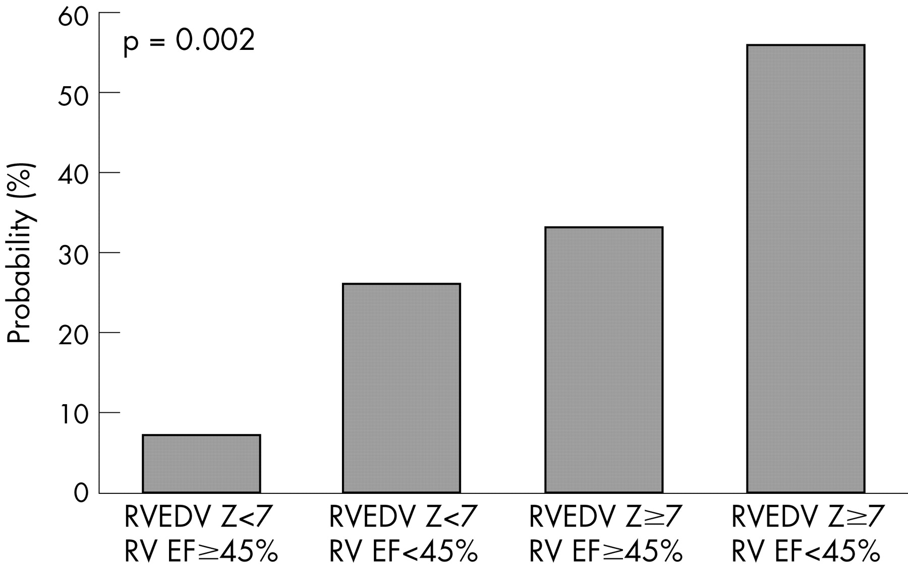
Multivariate analysis, controlling for length of follow-up from baseline to most recent evaluation, identified RV end-diastolic volume Z ⩾7 (OR = 4.55, 95% CI 1.10 to 18.8, p = 0.037) and LV ejection fraction <55% (OR = 8.05, 95% CI 2.14 to 30.2, p = 0.002) as independent predictors of major adverse clinical outcomes, with an area under the ROC curve of 0.850 (table 2). Restricting the analysis to RV parameters found that RV ejection fraction <45% (OR = 5.6, 95% CI 1.47 to 21.2, p = 0.011) and RV end-diastolic volume Z ⩾7 (OR = 4.00, 95% CI 1.10 to 14.6, p = 0.036) were independent predictors of major adverse clinical outcomes, with an area under the ROC curve of 0.807. Figure 1 shows the probability of a major adverse outcome in this model. The use of RV ejection fraction corrected for PR as suggested by Vliegen et al12 resulted in a weaker multivariate prediction model, with an area under the ROC curve of only 0.665.

Compared with the 70 patients who were alive and did not have a major adverse clinical outcomes, the four patients who died suddenly were older at the time of TOF repair (median age 18.1 vs 2.1 years, p = 0.006), more often had their repair before 1970 (75% vs 10%, p = 0.007), were older at the time of baseline CMR (median age 46.4 vs 20.9 years, p = 0.014), and tended to have a longer QRS duration (189 (18) vs 153 (26) ms, p = 0.064).
Separate analyses of outcomes in patients with RV outflow patch and those with RV-to-pulmonary artery conduit found similar predictors of major adverse clinical outcomes. Table 3 shows the trade-offs between sensitivity and specificity for different cut-off values of selected predictors of major adverse clinical outcomes. As expected, choosing lower cut-off values of RV ejection fraction or higher values of RV end-diastolic and end-systolic volumes resulted in increased specificity but decreased sensitivity for prediction of the primary outcome.
Factors associated with secondary outcomes
During follow-up, 27 patients (31%) underwent PVR. Indications for PVR included severe chronic PR associated with severe RV dilatation, RV dysfunction, symptoms and associated residual lesions (eg, tricuspid regurgitation), often in combination. Compared with those who did not undergo PVR, patients who did had a higher mean (SD) PR fraction (39 (16)% vs 28 (19)%; p = 0.01) and volume (22 (16) vs 14 (13) ml/m2; p = 0.026), more severe RV dilatation (RV end-diastolic volume Z-score 5.46 (3.65) vs 3.22 (2.86); p = 0.009), and lower LV end-diastolic volume Z-score (–1.08 (2.54) vs 0.43 (2.05); p = 0.011). They were more frequently receiving diuretics (30% vs 7%; p = 0.007) and were more often in NYHA class ⩾II (67% vs 38%; p = 0.032). Multivariate analysis, controlling for length of follow-up from baseline to PVR, identified PR fraction ⩾30% (OR = 4.79, 95% CI 1.44 to 16.0, p = 0.011) and diuretics at baseline (OR = 5.54, 95% CI 1.27 to 24.2, p = 0.023) as independent factors associated with PVR with an area under the ROC curve of 0.769.
An implantable defibrillator was placed in 20 patients (23%) during follow-up. Indications included a positive ventricular stimulation study, VT and syncope, often in combination. Compared with patients who did not have an implantable defibrillator, those who did were significantly older at the time of TOF repair (median age 8 vs 2.1 years; p = 0.044), had a lower RV ejection fraction (39 (12)% vs 50 (11)%; p = 0.001), higher RV end-systolic volume index (80 (35) vs 61 (31) ml/m2; p = 0.033), lower LV ejection fraction (56 (8)% vs 61 (9)%; p = 0.028), and longer QRS interval duration (172 (24) vs 154 (29) ms; p = 0.009). They were more frequently receiving diuretics (30% vs 9%; p = 0.025) and were more often in NYHA class ⩾II (60% vs 43%; p = 0.029). Multivariate analysis, controlling for the length of follow-up from baseline to the most recent evaluation, identified RV ejection fraction <45% (OR = 3.54, 95% CI 1.12 to 11.2, p = 0.031) and diuretics at baseline (OR = 5.25, 95% CI 1.15 to 24.1, p = 0.033) as independent factors associated with implantable defibrillator with an area under the ROC curve of 0.804.
Predictors of no clinical progression
Of the 88 cohort patients, 27 (31%) had no clinical worsening during follow-up. Patients who remained stable during follow-up underwent TOF repair at a younger age (median 1.7 vs 4.6 years; p = 0.008), had a higher RV ejection fraction (51 (11)% vs 46 (13)%; p = 0.037), and tended to have a higher LV ejection fraction (62 (9)% vs 58 (9); p = 0.065). The only independent predictor of “no clinical progression” was age at TOF repair <3 years (OR = 3.77, p = 0.013; area under the ROC curve 0.712).
DISCUSSION
This study found that RV dilatation and systolic dysfunction or LV dysfunction measured by CMR were independent predictors of major adverse clinical outcomes at a median of 21 years after TOF repair. Despite a relatively short follow-up period (median 4.2 years), patients were at significant risk of death, sustained VT, or functional deterioration if the RV was severely dilated (end-diastolic volume Z ⩾7) or had global systolic dysfunction (ejection fraction <45%).
Clinical implications
Previous studies on the risk of death or sustained VT late after TOF repair have demonstrated three major categories of outcome predictors: (a) history (syncope,17 older age at repair3); (b) electrophysiological markers (prolonged QRS duration,3 17 18 positive programmed ventricular stimulation study17); and (c) haemodynamic sequelae (cardiomegaly17 18). Most of these studies, however, lacked the tools to accurately assess ventricular mechanics, especially RV size and function. The use of CMR in the present study provided quantitative data on right and left ventricular size and function and PR—information that allowed assessment of their contribution to the risk of adverse clinical outcomes late after TOF repair. The finding that severe RV dilatation and even mild global RV systolic dysfunction place patients late after TOF repair at increased risk for adverse clinical outcomes resembles the experience with severe chronic aortic regurgitation. In that condition, it has long been recognised that depressed LV systolic function measured by ejection fraction is a strong predictor of poor outcome and, more recently, severe LV dilatation with normal systolic function has been recognised as a risk factor.19–21 In late survivors of TOF repair, both RV and LV mechanics might be impaired.4 6 The close relationship between RV and LV ejection fractions found in the present study and the observation that each predicts adverse clinical outcomes provide strong evidence that RV–LV interaction is key to understanding the pathophysiology that ultimately leads to poor outcomes late after TOF repair.
As expected, PR fraction and volume were closely associated with recommendation for PVR in this cohort. However, similar to the findings of our previous cross-sectional study,4 PR fraction or volume at a single time point was not predictive of major adverse clinical outcomes. Although PR is the primary source of chronic RV volume overload in these patients and correlates closely with RV size,22 the results of this study demonstrate that major adverse clinical outcomes relate primarily to effects of PR on the RV myocardium—dilatation and dysfunction.
This study was not designed to evaluate the benefits of PVR. The findings, however, provide important new information about the potential risks in adult patients with repaired TOF and chronic PR. Given the high incidence of major adverse clinical outcomes during a relatively short follow-up period and their relation to RV dilatation and dysfunction, further research is warranted to evaluate the potential benefits of PVR in patients with the risk factors found in this study, regardless of the presence of overt symptoms. Although there is no published evidence of a survival benefit from PVR, there is growing evidence of short-term benefits, including a decrease in RV size and improved exercise tolerance.12 13 23 24 Vliegen et al demonstrated significant improvement of RV size and function in patients referred for PVR before the onset of severe symptoms.12 In contrast, there is evidence that delaying PVR until the RV is severely dilated and poorly contracting often results in lack of functional recovery.7 11 25 Several groups recently published criteria for PVR in patients with repaired TOF.13 25–27 In view of the increased risk of poor clinical outcomes in these patients, the findings of the present study provide further support for not delaying PVR until RV failure and overt symptoms develop.
Cardiac magnetic resonance
This investigation demonstrates the clinical utility of CMR in outcome prediction and risk stratification for patients with repaired TOF. We chose to adjust RV and LV end-diastolic volumes to body size based on Z-scores to overcome differences between volume measurements by gradient echo cine (the technique used during baseline evaluation) and steady-state, free precession cine (current technique). Alfakih et al showed that compared with segmented gradient echo cine, LV and RV end-diastolic volumes measured by steady-state, free precession cine are ∼10% higher, LV mass is ∼17% lower, and RV and LV ejection fractions are not significantly different.28 The use of Z-scores to express the relation of an observed measurement relative to a sample of normal values minimises the effect of differences between image acquisition techniques. In this study, an RV end-diastolic volume Z-score of 7 corresponds to 172 ml/m2 in women and 185 ml/m2 in men using a steady-state, free precession cine technique.16
Study limitations
The study cohort consisted of consecutive patients enrolled during a period when CMR was transitioning from a targeted to routine test in patients with repaired TOF. Taken together with exclusion of patients with pacemakers or implanted defibrillators, the cohort may not accurately reflect the entire cross-section of this population.
CONCLUSIONS
In this cohort of late survivors of TOF repair, severe RV dilatation and either LV or RV dysfunction assessed by CMR predicted major adverse clinical events. This information may guide risk stratification and therapeutic interventions.
REFERENCES
Footnotes
Competing interests: None.
Funding: This work was supported in part by the National Institutes of Health (NIH/NHLBI 1P50 HL074734–01; Drs Geva, Powell and del Nido)