Article Text

Abstract
Background The presence of myocardial fibrosis by cardiac MRI has prognostic value in cardiac sarcoidosis, and localisation may be equally relevant to clinical outcomes.
Objective We aimed to analyse cardiac damage and function in detail and explore the relationship with clinical outcomes in patients with cardiac sarcoidosis using cardiac MRI.
Methods We included 81 consecutive patients with cardiac sarcoidosis undergoing cardiac MR. Left ventricular mass and fibrosis mass were calculated, and localisation was analysed using a 17-segment model. Participants underwent follow-up through 2015, and the development of major adverse cardiac events including ventricular tachyarrhythmias was recorded.
Results Increased left ventricular fibrosis mass was associated with increased prevalence of ventricular tachyarrhythmias (p<0.001). When localisation was defined as the sum of late gadolinium enhancement in the left ventricular basal anterior and basal anteroseptal areas, or the right ventricular area, it was associated with ventricular tachyarrhythmias (p<0.001). Kaplan-Meier analysis during a median follow-up of 22.1 months showed that both the mass and localisation groupings for fibrosis were significantly associated with major adverse cardiac events or ventricular tachyarrhythmias and that when combined, the risk stratification was better than for each variable alone (p<0.001, respectively). By Cox-proportional hazard risk analysis, the localisation grouping was an independent predictor for the both.
Conclusions In patients with cardiac sarcoidosis, both fibrosis mass and its localisation to the basal anterior/anteroseptal left ventricle, or right ventricle was associated with the development of major adverse cardiac events or ventricular tachyarrhythmias. Cardiac MR with late gadolinium enhancement may be useful for improving risk stratification in patients with cardiac sarcoidosis.
- MYOCARDIAL DISEASE
This is an Open Access article distributed in accordance with the Creative Commons Attribution Non Commercial (CC BY-NC 4.0) license, which permits others to distribute, remix, adapt, build upon this work non-commercially, and license their derivative works on different terms, provided the original work is properly cited and the use is non-commercial. See: http://creativecommons.org/licenses/by-nc/4.0/
Statistics from Altmetric.com
Key questions
What is already known about this subject?
The presence of myocardial fibrosis by cardiac MRI has prognostic value in cardiac sarcoidosis.
What does this study add?
The fibrosis localisation to the basal anterior/anteroseptal left ventricle, or right ventricle is associated with the development of major adverse cardiac events or ventricular tachyarrhythmias.
How might this impact on clinical practice?
Cardiac MRI with late gadolinium enhancement is promising for predicting adverse events including ventricular tachyarrhythmias.
Introduction
Cardiac involvement in sarcoidosis occurs in 20–30% of patients in autopsy studies,1 ,2 and has been closely associated with prognosis.3 ,4 A previous study reported that sudden cardiac death (SCD) occurred in two-thirds of patients with histological evidence of cardiac sarcoidosis (CS).5 Ventricular tachyarrhythmia (VA), resulting from myocardial granulomas and fibrosis that cause electric instability, has been identified as the underlying mechanism of SCD. The 5-year mortality rate in patients with CS when structural or functional abnormality is present, according to the diagnostic criteria of the Japanese Ministry of Health and Welfare (JMHW), has been reported at 40%, with the cause of death being the progression of heart failure (HF) or VA.6 However, the factors related to the clinical manifestations and outcomes of CS remain unclear and the risk stratification and clinical management are difficult, particularly for VA and SCD.
Late gadolinium-enhancement cardiac MR (LGE-CMR) has been used to detect myocardial damage/fibrosis, and recent findings have suggested that it is more sensitive than clinical diagnostic criteria for detecting CS.7 ,8 A recent study demonstrated that LGE-positive patients had a higher rate of adverse events (a Cox HR of 31.6 for death, aborted SCD, or appropriate ICD discharge), compared with LGE-negative patients in a cohort of patients with suspected CS.9 Although myocardial fibrosis indicated by LGE may predict potentially lethal events, the utility of LGE in patients with sarcoidosis who present with high-grade cardiovascular symptoms, such as HF, VA and advanced atrioventricular block (AVB), has not yet been established. In addition, detailed risk stratification in patients with CS using LGE-CMR remains challenging. Therefore, we analysed cardiac damage and function in detail and explored the relationship with clinical outcomes including VA, using CMR in patients with CS. We assumed that not only fibrosis mass but also its localisation might help to identify the risk of clinical outcomes in patients with CS.
Methods
Study protocol
We enrolled consecutive patients with CS referred for CMR between 2008 and 2015 at three referral centers (Kindai University Hospital, Osakasayama, Japan; Kitano Hospital, Osaka, Japan; and Shiga Medical Center for Adults, Moriyama, Japan). The study was approved by the Institutional Review Boards.
Patients were enrolled if they fulfilled the following criteria: (1) definite CS: CS diagnosed by the 2006 revised JMHW criteria;10 or (2) suspected CS: strongly suspected CS based on a constellation of clinical characteristic manifestations and positive findings on echocardiography, 18F-fluorodeoxyglucose-positron emission tomography (FDG-PET), or CMR, with or without extra-CS, and after the exclusion of other known cardiac diseases.11 The characteristic positive findings of CMR and FDG-PET were based on those detailed in previous reports.7 ,12
CMR image acquisition
CMR was performed using a 1.5 T scanner (Intera 1.5T; Philips Medical Systems, the Netherlands), according to a standardised protocol. Cine images were acquired with a steady-state free-precession breath-hold sequence in three long-axis planes and contiguous short-axis slices (10 mm, no gap) from the atrioventricular ring to the apex. The visualisation of myocardial oedema was performed using a T2-weighted triple inversion recovery breath-hold pulse sequence. LGE images were obtained 10–15 min after intravenous administration of 0.2 mmol/kg gadolinium-DTPA (Magnevist; Bayer Schering Pharma, Germany) using an inversion recovery gradient echo sequence with 5 mm slice thickness at the same position as the long-axis and short-axis cines at end-diastole. The inversion time was adjusted per patient to an optimal null signal from a normal myocardium, typically between 250 and 350 ms.
Image analysis
LV volume, LV mass (LVM), and wall thickness were calculated with commercially available workstations (Aze Virtual Place; Aze , Japan) as previously described.13 The endocardial and epicardial borders of the LV myocardium were manually planimetered on successive short-axis cine images at end-diastole and LVM was derived by the summation-of-the-disks method and multiplication of the myocardial muscle volume by 1.05 g/cm3. To assess LGE, all short-axis slices from the base to the apex were visually inspected to identify areas of normal (completely nulled) myocardium. The mean signal intensity (and SD) was derived, and a threshold of 6 SD above the mean was used to define the areas of LGE.14 Summing the planimetered areas of LGE in all short-axis slices yielded the total volume (LV fibrosis mass; g), which was also expressed as a proportion of total LV myocardium (% LGE).
Local wall thickness, LGE, and T2-weighted images were also analysed based on the American Heart Association's (AHA) 17-segment model. To evaluate the relationship between localisation of LGE and clinical outcomes, a localisation score (LS) was defined as the summation of LGE enhancement in segments identified by LGE localisation analysis. All analyses were performed by an experienced specialist, and reviewed and confirmed by a second expert. Both independent observers were blinded to the patients' identities and clinical profiles. Any discrepancies in analysis were adjudicated by a senior observer.
Clinical follow-up
Long-term clinical follow-up was for 50 months after the CMR testing and accomplished by patient-completed questionnaire, telephone interview or chart review. Major advanced cardiac events (MACE) were defined as the combination of cardiac mortality, admission for worsening HF, advanced AVB and VA. VA was defined as >3 consecutive ventricular contractions, appropriate implantable cardioverter defibrillator (ICD) discharge or ventricular fibrillation (VF). Sustained ventricular tachycardia (VT) was defined as a ventricular rhythm faster than 100 bpm lasting at least 30 s. All events were based on clinical diagnosis but were validated by reviewing the patients' medical records. Advanced AVB, VA or sustained VT was also confirmed by ECG or ICD records.
Statistical analysis
As part of the univariable analysis for continuous variables, comparisons among groups were performed with student's t-tests, one way analysis of variance, and Mann-Whitney tests. Pearson χ2 and the Fisher's exact test were used to assess differences in categorical variables. Cut-off levels of LV fibrosis mass index for VA events and the sensitivities and specificities of the cut-off levels were calculated using a receiver operating characteristics (ROC) curve analysis. Event-free survival curves were analysed using the Kaplan-Meier method and comparison between curves was carried out using log-rank tests. The multivariate analysis of clinical outcomes was evaluated with Cox's proportional hazard model, using JMP V.10.0 (SAS Institute , USA). HRs and 95% CIs were calculated. A p value <0.05 was considered significant. All results are expressed as mean±SD.
Results
Baseline patient characteristics
In total, 81 consecutive patients were enrolled, of which 35 were diagnosed as definite CS using the 2006 revised JMHW criteria and 46 were diagnosed as suspected CS clinically (table 1). Furthermore, 38 (46.9%) patients had extracardiac involvements and 43 (53.1%) patients were diagnosed as having isolated CS, with 31 (38.3%) patients having prior VA events, 20 (24.7%) having documented advanced AV block, and 24 (29.6%) being complicated by HF. Plasma B-type natriuretic peptide (<18.4 pg/mL) and ACE inhibitors levels (<21 IU/L) were increased in 84.0% and 32.9% of the patients, respectively. Although just 15 (18.5%) patients had taken corticosteroids at baseline, a further 27 (33.3%) had taken them during follow-up.
Baseline clinical characteristics and CMR findings
CMR findings and VA events
In the CMR examination at baseline, a reduced EF (<50%) was observed in 58.8% and LGE was present in 94.9%, with lesions frequently observed in the basal septum (Segment 2: 75.9%; Segment 3: 65.8%) and RV (57.0%). The mean % LGE and FM index were 16.6%±12.8% and 11.3±9.6 g/m2, respectively. During a median follow-up of 22.1 months, 25 (30.7%) patients had VA episodes of which 8 (9.9%) where new onset and the other 17 (21.0%) where recurrences. Any VA event was observed in 39 (48.1%) patients.
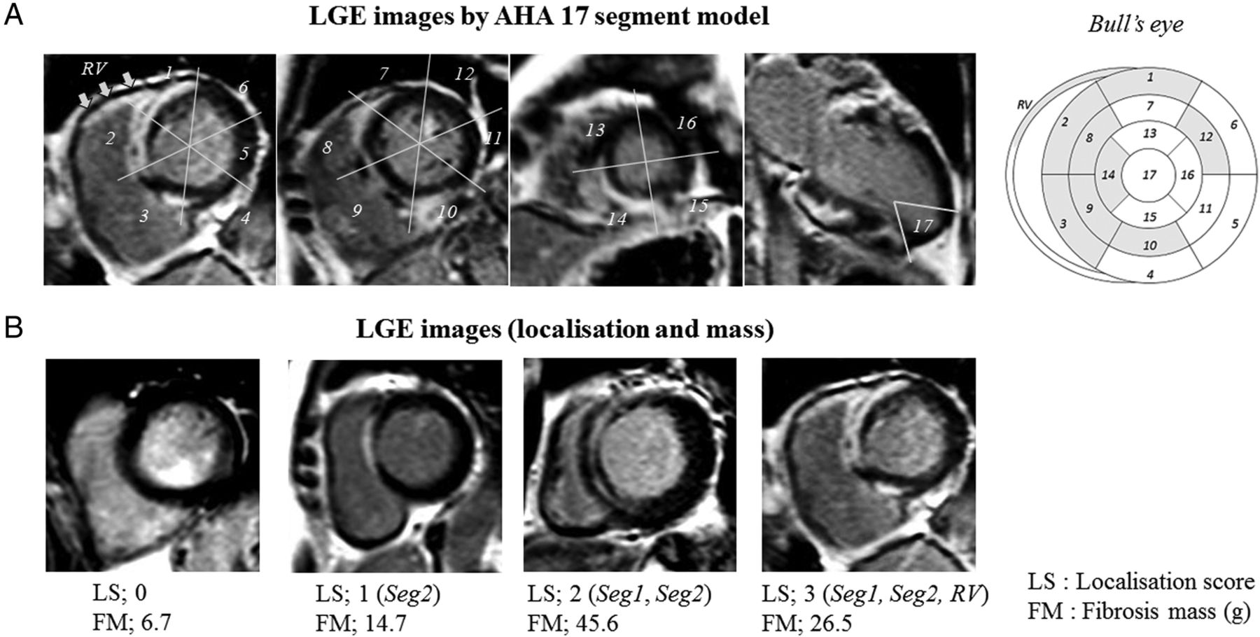
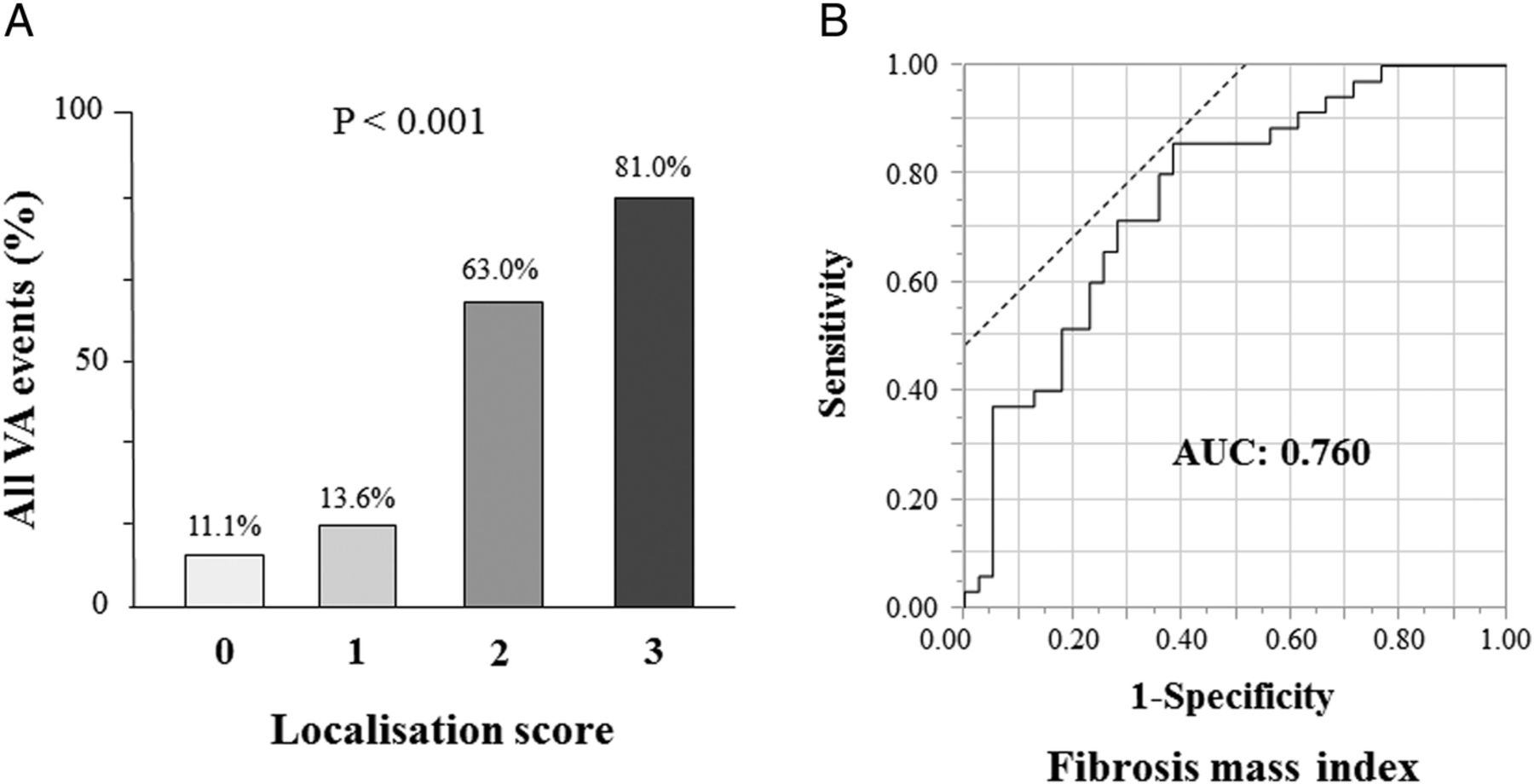
As shown in table 2, all VA events (ie, a history of VA and new-onset VA events) were highly associated with the existence of LGE in the basal anterior (segment 1) and basal anteroseptal (segment 2) LV, as well as the RV (p<0.01). Among patients with VA, almost all had LGE in segment 2 and LGE was more frequently present in segment 1 and the RV than in patients without VA. Accordingly, we defined the LS as the summation of the LGE in segment 1, segment 2, and the RV, with a potential score of 0 to 3. Figure 1 shows representative LGE images according to the AHA's 17-segment model and representative cases stratified by the LS. As shown in figure 2, when patients with all VA events were stratified by LS, LS was significantly associated with all VA events (p<0.001). Increased LV FM index was also associated with all VA events (p<0.001), and ROC analysis showed that the area under the ROC curve (AUC) for FM as an indicator of all VA events was 0.760 (95% CI 0.651 to 0.870) (figure 2). The optimal cut-off value of LV FM index was 5.12 g/m2, with a sensitivity of 86% and a specificity of 62%.
Association of VA events with localisation of LGE according to the 17-segment AHA model

Representative LGE images using the AHA 17-segment model (A) and representative cases stratified by the LS (B). AHA, American Heart Association; FM, fibrosis mass; LGE, late gadolinium enhancement; LS, localisation score.

Relationship between LV fibrosis mass index or localisation score and VA events. Stratification of patients with VA events by localisation score (A). Receiver operating characteristic (ROC) curves for LV fibrosis mass index as an indicator of all VA events (B). AUC, area under the ROC curve; VA, ventricular tachyarrhythmia.
Clinical outcome and risk stratification
During a median follow-up of 22.1 months (lower quartile, 7.4 months; upper quartile, 38.1 months), 30 MACEs occurred: 3 patients died due to SCD or HF, 7 were admitted for worsening HF and 4 patients had advanced AVB. Twelve severe ventricular arrhythmias episodes such as sustained VT and VF occurred. In 10 patients, the origin was estimated by 12-lead ECG and 8 patients showed the ECG with inferior axis. They were from left/right ventricular outflow tract or high LV septum, which were associated with LS (the existence of LGE in segment 1, 2 or RV).
For the Kaplan-Meier analysis, patients were divided into two groups according to the optimal cut-off for LV FM index (<5.12 g/m2 (n=29) or ≥5.12 g/m2 (n=45)), or the LS (≤1 (n=32) or ≥2 (n=47)). Patients with an LV FM ≥5.12 g/m2 or an LS ≥2 showed significantly worse prognoses in terms of both MACE and VA events (figure 3). In sustained VT or VF events, only increased LS was associated with worse prognosis (p=0.006). Lower EF (<45%) was mildly associated with the worse prognosis in the MACE (p=0.045), and extracardiac sarcoidosis involvement was not associated with the both prognoses (p=0.330 and P=0.930, respectively).
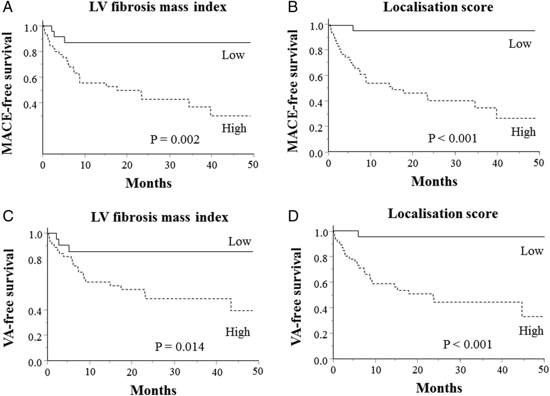
Kaplan-Meier analysis for the MACE-free (A and B) and VA-free survival (C and D) in patients stratified by baseline LV fibrosis mass index (A and C) and localisation score (B and D). Lower group, solid line; higher group, dotted line. MACE, major adverse cardiac event; VA, ventricular tachyarrhythmia.
In multivariate Cox proportional hazard analysis with age, gender and FM index, the LS was independently associated with MACE and VA events, as shown in table 3. When the FM index as a continuous variable and the LS as throughout its range were entered into the analysis, the LS was still an independent predictor for MACE and VA (P<0.001 and P=0.002, respectively). Furthermore, as shown in figure 4, the combined analysis for the higher FM index and LS groups showed that the combination was associated with the highest risk of MACE and VA events. Notably, all patients with admission for worsening HF or advanced AVB also belonged to this group. In contrast, patients with a FM index <5.12 g/m2 and an LS ≤1 had no MACEs, or no VA events.
Multivariate Cox proportional hazard analysis

Risk stratification of clinical outcomes by the combined analysis of LV fibrosis mass and localisation score: MACE (A), VA events (B) and all VA events (a history of VA and new-onset VA events) (C). MACE, major adverse cardiac event; VA, ventricular tachyarrhythmia.
Comparison between definite and suspected CS
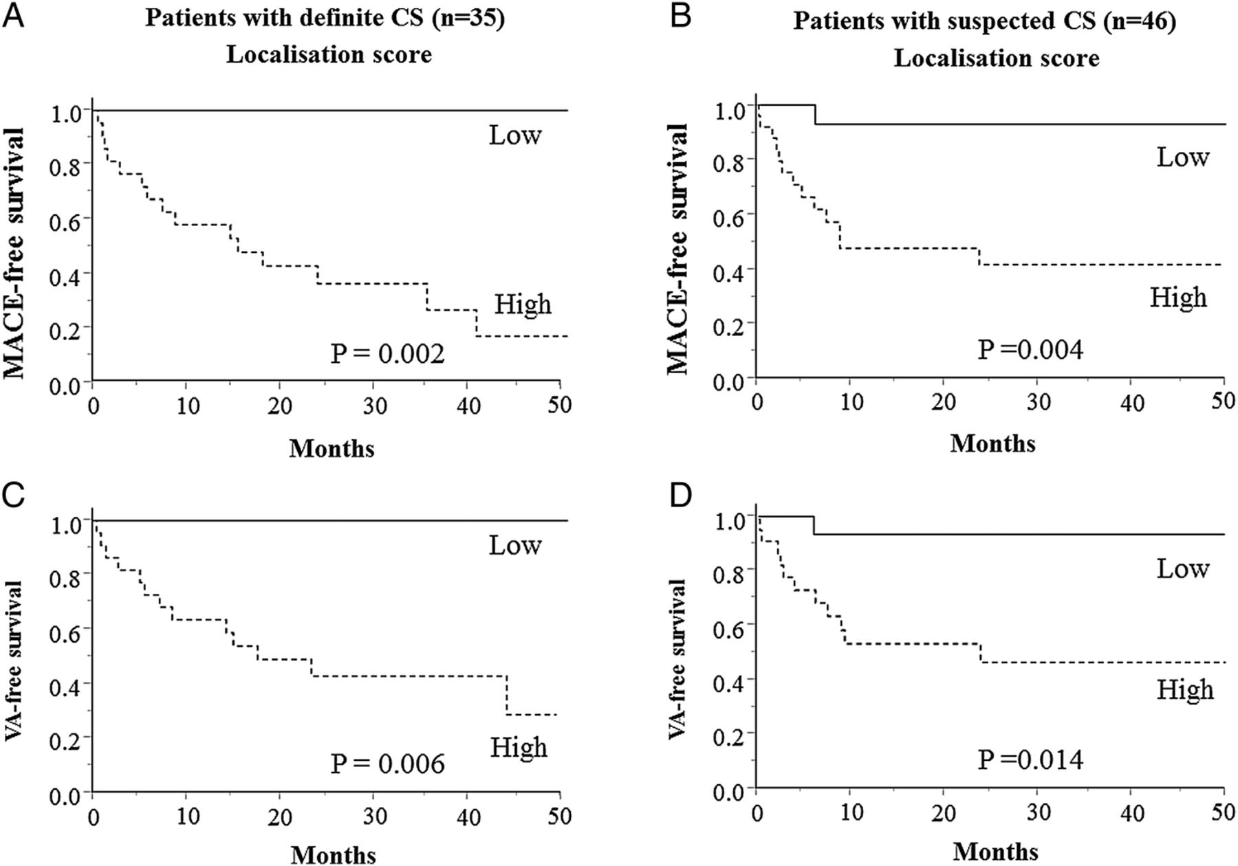
In comparison of baseline clinical characteristics between patients with definite and suspected CS, frequent extracardiac involvement and increased plasma ACE level were observed in definite CS (p<0.01, respectively) (see online supplementary table S1). Also, corticosteroid therapy was frequently performed (p=0.003). In CMR analysis, no significant differences were observed between them (see online supplementary table S2). In Kaplan-Meier analysis, each group also showed that the higher LS group (≥2) had a worse prognosis in both MACE and VA events (figure 5).
Kaplan-Meier analysis for the MACE-free or VA-free survival in patients with definite CS (A and C) or suspected CS (B and D) stratified by localisation score. Lower group, solid line; higher group, dotted line. CS, cardiac sarcoidosis; MACE, major adverse cardiac event; VA, ventricular tachyarrhythmia.
Supplementary tables
Discussion
The major findings of this study are as follows. First, VA was the most frequent cardiac event in CS, and VA events, including a prior history of VA, occurred in 48.1% of patients. Second, an LV FM index ≥5.12 g/m2 was shown to predict MACE and VA. Third, a developed LS, defined as the summation of the LGE in the basal anterior LV, basal anteroseptal LV and RV, was able to predict VA when ≥2. Fourth, the combination of both FM and LS improved the risk stratification for predicting clinical outcomes, including VA.
Prognosis and VA in patients with CS
In a retrospective autopsy series of SCD in sarcoidosis, 25 of 41 patients had evidence of cardiac involvement and 10 of 25 patients had no evidence of sarcoid in any other organ.15 Fleming et al16 reported that SCD occurred in 48 of 197 (24%) patients with CS in their cohort. In a retrospective study of Japanese patients with CS, sustained VT was shown to be an independent risk factor for mortality, suggesting that VA is very important for prognosis of CS.6 In the present study, only two patients had SCD caused by a VA, or 12 patients had severe ventricular arrhythmias. We therefore tried to identify a better risk stratification tool for VA in CS that was more closely associated with SCD.
In an autopsy series of SCD in sarcoidosis, the ventricular septum had the largest percentage of involvement and ∼50% of the cases were involved in the RV apex and septum.15 Kumar et al17 reported that among 21 patients with CS and VT, 16 of 18 patients had an RV scar and 14 of 15 patients had an LV scar, with LV scarring frequently present in the septal (11/15) and anterior (7/15) LV. Similarly, our findings indicated LGE in basal septum at a high frequency among the patients with VAs (segment 2, 92.1%; segment 3, 79.0%), followed by the RV (73.7%) and basal anterior LV (segment 1, 60.5%). In a recent cohort study of CS by Crawford et al,18 the presence of LGE in the RV was associated with adverse events, including VT/VF. Consistent with their studies, our results suggest that the presence of LGE in the RV may be important for the origin of VT.
For further risk stratification, we utilised a three-segment model (segment 1, 2 and RV), which showed highly significant differences in the presence of LGE between patients with and without VA (table 2). These were then used to define the LS, which was itself highly associated with not only all VA events (figure 2) but also follow-up VA events (P<0.001, data not shown). The reason the LS was useful for risk stratification in VA is unclear. However, myocardial scar has been known to be an origin and a specific substrate of VA and it is proposed that the scars in the His-Purkinje system, LV outflow-tract in the basal septum and the anterior wall may participate in VA generation or SCD processes in CS using voltage cardiac mapping.17 Indeed, in the present study, the origin of sustained VT was estimated in 10 patients and the 80% was from left or right ventricular outflow tract or high LV septum, suggesting the close relationship between VT origin and LS (ie, the existence of LGE in segment 1, 2 or RV). With regard to the importance of RV involvement, Schuller et al19 reported that RV dysfunction, in particular, was associated with appropriate ICD therapy in patients with CS. Further mechanistic studies for VA generation will clarify and improve the risk stratification in CS.
LGE and VA in cardiomyopathies
In studies of LGE in patients with VA, including both ischaemic and non-ischaemic cardiomyopathies, the presence of fibrosis was an independent predictor of adverse outcomes.20 In a meta-analysis by Kuruvilla et al21, the presence and extent of LGE were associated with increased risk of all-cause mortality, HF hospitalisation, and SCD in patients with overall non-ischaemic cardiomyopathy. However, given that the basic pathophysiology is different for each cardiomyopathy, the relationship between LGE and each event may also diverge, suggesting that LGE quantification cannot be uniformly adapted to all cardiomyopathies. In a cohort study of 1293 patients with hypertrophic cardiomyopathy, the extent of LGE was associated with an increased risk of SCD, with the adjusted HR reported as 1.46 per 10% increase in LGE.22 In a study of dilated cardiomyopathy, mid-wall fibrosis was present in 142 (30%) patients, and its assessment with LGE-CMR imaging provided independent prognostic information.23
Similarly, the presence of LGE among patients with suspected CS yielded a Cox HR of 31.6 for death, aborted SCD and appropriate ICD discharge, and of 33.9 for events including death and VT during a 3.4-year follow-up in a recent prospective study.9 However, they found no relationship between cardiac events including VA and LGE extent, the studied cohort consisted of a broad range of patients with systemic sarcoidosis, and the % LGE in all patients was lower than that in our study cohort (4.4% vs 16.6%, respectively). In the present study, our cohort may be representative of more advanced CS, resulting in the increased LV FM by quantification being significantly associated with MACE or VA events by Kaplan-Meier analysis, and the ROC curve analysis showing an optimal cut-off value of 5.12 g/m2 in LV FM index for VA events. A recent study in 43 Japanese patients with CS taking steroid therapy, where % LGE was 19%±10% (5-SD method), showed that large-extent LGE (ie, % LGE ≥20%) was independently associated with combined adverse outcomes.24 Crawford et al18 also reported that LGE involving ≥9 segments in a combined left and right ventricular segmental analysis resulted in a 92% sensitivity and 88% specificity for differentiating patients with VT/VF from those without VT/VF (AUC, 0.90). In addition, the results of the present study suggest an incremental effect of the LS on LGE extent (LV FM) by LGE assessment, and that a combined analysis of LS and LV FM may be better for the risk stratification of clinical outcomes in CS.
Study limitations
Several limitations should be considered when interpreting our results. First, the study population was relatively small and could have resulted in low statistical power. However, the size of our cohort was average relative to that of other studies of CMR in CS.17 ,18 Also, the retrospective nature of the study may suggest some impact of the missing or insufficient data on the present results.
Second, about half of the patients did not fulfil the JMHW diagnostic criteria (suspected CS), most of whom had isolated CS. Recently, several reports have suggested that there are similar clinical characteristics and clinical outcomes for suspected CS that does not fulfil the JMHW diagnostic criteria but that satisfies only the guidelines for clinical cardiac diagnosis.25 To test this, we analysed and compared the data between the patients who did (definite CS) and did not fulfil the JMHW diagnostic criteria (suspected CS), and found that the clinical characteristics were indeed similar and that the higher LS group (≥2) had a worse prognosis in each group (figure 5).
Third, the study population consisted of patients with CS who could undergo CMR. In particular, most patients with previously implanted cardiac devices and those with severe renal failure were not included in the present study since they could not tolerate the procedure. Therefore, the event rate and clinical characteristics might be underestimated in the present analysis.
References
Footnotes
MY and YI both authors contributed equally to this work.
Contributors MY and YI are the primary investigators. TK, TI, YI, TN, TK, YM are the secondary investigator (data sampling/analysis). SI, M I, TK, SM are consultants and supervisors.
Competing interests None.
Ethics approval Kindai University Faculty of Medicine.
Provenance and peer review Not commissioned; externally peer reviewed.